Pigs are equipped with one of the most powerful olfactory (sense of smell) systems in the animal kingdom, with over 8,000 scent receptors in their nose. This remarkable ability not only aids in detecting a vast array of scents but also plays an essential role in their survival, behaviour and social interactions.
The Science Behind Pig Olfaction
At the core of this acute sense of smell are the cells in the nasal epithelium. These cells convert incoming odour molecules into electrical signals, which are then transmitted via the olfactory nerve to the brain. This process allows pigs to detect and interpret a wide range of scents.
Additionally, pigs have a specialised Vomeronasal Organ (VNO) located in their upper air passages. This organ contains receptors that have direct access to the central nervous system (CNS) through the accessory olfactory bulb, allowing pigs to process pheromones, which play a vital role in their social and reproductive behaviours.
An Acute Sense of Smell
In a test situation, sows were able to distinguish between identical cards they had previously touched and those they had not. Even after the cards were washed, the sows could still recognise the odour they had deposited on them hours earlier. This shows how powerful the pigs’ memory and sense of smell truly are.
More Than Just a Nose—A Versatile Tool
In addition to housing their vast array of scent receptors, the pig’s nose serves another critical function: it’s also their primary digging tool. Pigs instinctively root through the ground to find food, using their strong snouts to dig into the soil. This behaviour, known as foraging or rooting, allows them to locate roots, tubers, and insects that lie beneath the surface. Their noses are not only finely tuned to detect scents, which is up to 7 miles, but also physically built for digging and exploring their environment.
The Role of Smell in Pig Behaviour
Pigs use olfactory cues in various aspects of their behaviour:
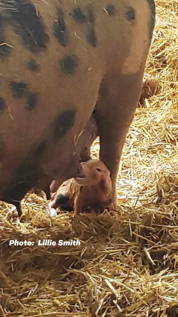
- Piglets rely on scent to recognise their mother and locate the correct teat position.
- Older pigs use smell as the primary basis for individual recognition within their social groups.
- Reproductive behaviour is largely influenced by scent. The presence of a mature boar’s scent can trigger estrous behaviour in gilts. Boar saliva and urine contain a steroid hormone called androgen, which acts as a pheromone to elicit this response.
Fascinatingly, the same androgen compound is found in the blood and urine of young pigs and has been shown to reduce aggressive behaviours when applied to other pigs. And interesting study proving the importance of how chemical communication is to maintaining social harmony within pig groups.
Conclusion
With over 8,000 sensors and a strong nose built for digging, pigs’ olfactory systems are integral to their behaviour, survival and interaction with their environment. Their extraordinary sense of smell enables them to engage in complex social behaviours, manage reproductive cues and even use their snouts to forage for food. Understanding these abilities can provide valuable insights into their welfare and management, especially with breeding programs. Whether recognising a scent long after it was left or foraging beneath the earth, the pigs’ nose is truly one of the most powerful and sensitive tools.

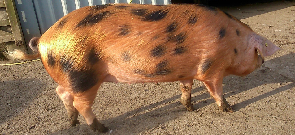
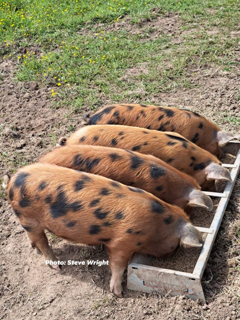
The Oxford Sandy and Black Pig Group (OSBPG) is the UK’s only pedigree pig breed registered as a charity in both England & Wales (1190469) and Scotland (SCO52662). We are dedicated to creating a better future for the breed by preserving bloodlines and enhancing breeding potential. In partnership with Independent Pork Producers, Breeders, and Oxford Sandy and Black Keepers and their families, we work to strengthen the breed’s legacy. Your support is invaluable – please click the donate button to help us continue our efforts and provide resources and initiatives that drive our mission forward.
Follow us on Facebook and see how we support, help and inspire individuals about our beautiful rare breed the Oxford Sandy and Black Pig