CANNIBALISM IN PIGS


Understanding and Managing Cannibalism in Pigs

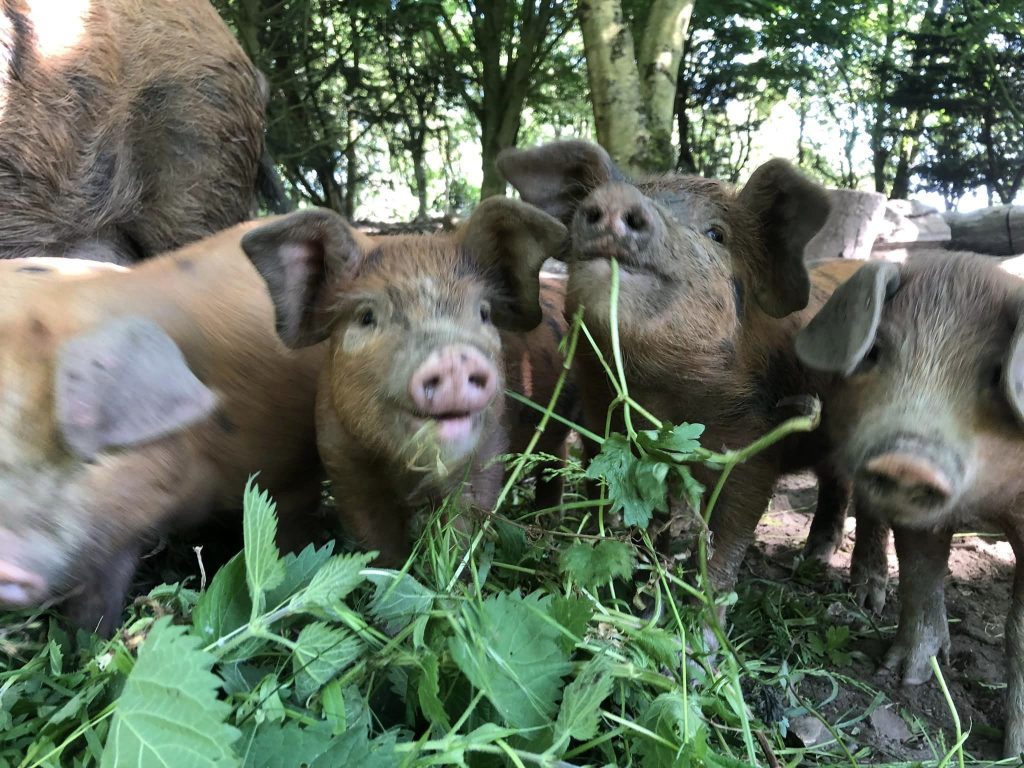
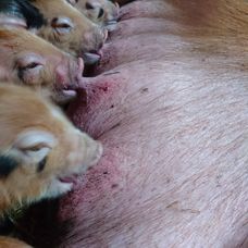
In recent months, the Oxford Sandy and Black Pig Group (OSBPG) have seen reports that have surfaced regarding sows and occasionally gilts exhibiting cannibalistic behaviour towards their piglets. These incidents typically occur in the context of large litters, ranging from 12 to 15 piglets, and can happen in both outdoor and indoor farrowings. Despite providing ample space, good lighting, and regular feeding, the sows tend to reduce the litter size to 7 or 9 piglets, indicating a contentment with this smaller size.

The phenomenon of cannibalism in pigs raises questions about its underlying causes.

Is it due to inherent aggression in certain pigs, close breeding, or perhaps a natural response indicating an inability to care for all piglets until weaning age? While some attribute it to shortcomings in husbandry skills, research suggests that cannibalism, termed “savaging,” is a recognised but poorly understood behaviour in pig farming.

Genetic Factors: Savaging is observed across various mammal species, including wild boars, and appears to be more prevalent in species with multiple births. While there are differences in general aggression among domestic pig genotypes, savaging does not necessarily run in families or result from inbreeding. Anecdotal evidence hints at certain commercial genotypes being more predisposed to savaging than others.

Predisposing Factors: Several factors tthat may predispose sows and gilts to exhibit cannibalistic behaviour:

  • Poor farrowing environment
  • Lack of empathy and inadequate handling
  • Possible effects of feed intake (lower intake before farrowing leading to hunger)
  • Pain during farrowing
  • “Storms” triggered by other savaging females in the same farrowing room
  • Inefficient cross-fostering practices

Management Strategies: Addressing cannibalism requires a multifaceted approach which may be considered such as:

  • Regular and gentle handling of pregnant gilts, emphasising empathy
  • Providing sufficient space for gilts at least a week before farrowing in indoor settings
  • Offering bran or grass before farrowing to promote gut health
  • Encouraging nest-building behaviour by providing ample straw pre-farrowing
  • Playing background music during the farrowing period may have a calming effect
  • Identifying problem animals early based on behavioural cues and considering treatments – with advice taken from your Veterinarian
  • Confining piglets to the creep area at the first sign of trouble during farrowing
  • Avoiding cross-fostering litters onto gilts
  • Consider culling sows that exhibit repeated savaging behaviour across multiple litters

In conclusion, cannibalism in pigs remains a complex and challenging issue in pig farming, necessitating a combination of proactive management practices and vigilant observation to mitigate its occurrence and impact on litter survival rates.


The Oxford Sandy and Black Pig Group is UK’s only pig breed that is a registered charity in England & Wales (1190469) and Scotland (SCO52662). We are creating a better future for our breed the bloodlines and it breeding potential together with our Independent Pork Producers, Breeders and Oxford Sandy and Black Keepers and their families. Please click the donate button so we may continue to look after our breed and our supporters

Follow us on Facebook and see how we support, help and inspire individuals about our rare breed Oxford Sandy and Black Pig

Coccidiosis

Coccidiosis affects all ages of pigs and is a tacky or watery diarrhoea, piglets do not seem to grow very well and can cause death in piglets. It stems from poor hygiene.

Causes

Coccidia are single cell protozoan parasites which multiply in the cells lining the intestine. Cystoisospora suis is the main pathogen of piglets and is responsible for the most coccidial diarrhoea. Cryptosporidium parvum, C. suis and other genotypes may also affect piglets but occurs commonly in weaned pigs and Eimeria species occur in weaned pigs, finishers and gilts but not in piglets. The resistant oocysts of Cystoisospora. suis are eaten and reach the upper small intestine where small sporozoites hatch and invade cells of the intestinal lining within 24 hours. They multiply asexually, damaging their host cell, to give rise to first stage merozoites and then to a further two stages within cells of the small intestinal lining. Each fourth stage cell reaches the lower part of the small intestine at about 4-5 days after infection and invades an epithelial (lining) cell to produce the sexual stage. Some become groups of microgametes and some remain single to become macrogametes. Mating in the gut produces oocysts which appear in the faeces as oocysts within 5 days of infection. Eimeria species multiply in the same way as C. suis, but Cryptosporidium remains in the absorptive, apical part of the cell and appears to prevent absorption of nutrients and causes cell death.

Transmission

Coccidia are passed out in the faeces and Cystoisospora suis oocysts develop within 48 hours at 24-27ºC into resistant hard walled sporulated oocysts which are infective. They are resistant to drying and can persist in piggeries and soil for up to 10 months. The major source of infective oocysts is the faeces of piglets aged between 2 and 4 weeks, when from 4,000 to 400,000 oocysts per gram may be present. Sows may pass a few oocysts and infect their litters, but the major source of both direct and indirect infection is the piglet. Transmission of Eimeria species resembles that of Cystoisospora, although the age range affected differ, but some Cryptosporidium species have more than one host species. The route of infection is oral in all cases.

Signs

Coccidiosis causes mal-absorption, diarrhoea, loss of condition and some deaths. The faeces of affected piglets may be whitish and firm, there may be transient pasty diarrhoea or profuse yellowish, watery diarrhoea between 5-15 days of age and most commonly from 7-10 days of age. Affected piglets may be thinner than unaffected litter mates and remain gaunt and hairy for some weeks thereafter. Severely affected piglets may die. Mortality rates may reach 20%. The diarrhoea responds poorly to antimicrobial therapy and slow growth occurs in recovered animals and diarrhoea rarely continues beyond weaning. Wasting and diarrhoea have been recorded in gilts, finishing pigs and wild boar infected with Eimeria spp. In experimental E. neodebliecki infection, frothy, mucoid diarrhoea occurred 9-12 days post infection. Most infections with C. parvum are asymptomatic (90% in one study). Clinical signs rarely appear before 3 days of age and are most commonly reported in piglets at 10-21 days of age. Affected piglets are depressed and pass a watery, sometimes brownish, diarrhoea, which may persist for 3-5 days. The organism can be recognised in the faeces of diarrhoeic weaners. Affected weaned pigs are often depressed and anorexic, in poor condition with a rough dirty coat and pasty faeces.

Treatment and prevention

Coccidiosis in piglets can be treated with trimethoprim sulphonamide and they should be supported with electrolyte. Toltrazuril given as an oral suspension by stomach tube on day 3 prevents diarrhoea, prevents oocyst shedding and can maintain piglet growth. Scrupulous attention should be given to hygiene in control, as the high levels of oocysts passed by piglets and their ability to survive for months make environmental contamination important. Sows should be cleaned on entry to the farrowing houses which should be disinfected or fumigated with methyl bromide or ammonia (final concentration 2%). Steam cleaning at 65˚C for 15 minutes and proprietary disinfectants can also be used to destroy oocysts. Less disease and oocyst shedding has been demonstrated on fully slatted cleaned floors. Trimethoprim sulphonamide may be given by injection or orally to older pigs with Eimeria species and the inclusion of an anticoccidial such as amprolium at 1 kg permix per tonne in sow feed for 7-10 days prior to farrowing and for 2 weeks afterwards will reduce the shedding of oocysts by sows and other pigs. No treatment has yet been shown to affect Cryptosporidia. The oocysts can be destroyed by the same disinfectants as those of the other coccidia.

Be aware

Cystoisospora and Eimeria infections are of no public health importance, but the pig may be a source of infection with Cryptosporidium for humans and vice versa.

Biotin Deficiency – Nail Growth

This affects all age groups from growers, gilts, sows and boars. It is caused by diet and poor floors and effects Lameness, claw defects, poor growth, diarrhoea, hair loss, scaly skin, small litters.

Causes

Dietary biotin (a water-soluble B vitamin) is normally present in adequate amounts in pig rations, although in feeds containing wheat or barley, the vitamin may be less readily available to pigs than in maize-based diets. Biotin is a cofactor in a number of body enzymes especially in the carboxylation (a chemical reaction in which a carboxylic acid group is produced by treating a substrate with carbon dioxide) or transcarboxylation reactions required for energy metabolism. In growing pigs lipogenesis (fat formation) is affected when the important enzyme, Acetyl CoA carboxylase cannot be formed. Deficiency in biotin on wheat and barley-based rations occurs quite commonly and at least 180 mg/tonne should be present for health.

Mode of transmission

Biotin deficiency is not infectious and occurs where rations are deficient in the vitamin.

Clinical signs

Early changes in biotin deficiency are slight but include progressive hair loss, dry and scaly skin and a white film and transverse grooves on the tongue. After 5-7 weeks on a deficient diet, claw defects occur. Erosion of the heel occurs first and is followed by cracking of the sole. Cracks appear in the now rubbery horn of the sole and the claw wall and result in lameness. Secondary infection may occur. In most cases, foot lesions in sows appear on the underside of the hoof and are only seen when the claw is cleaned. Others appear as longitudinal cracks on the wall originating in the coronary band. Heel and sole erosions and cracks also occur. Inspection of the sows’ feet after thorough cleaning will allow the claw defects to be seen clearly. Cracks arising from the coronary band and erosions of the sole and heel and the presence of alopecia and scaly skin suggest biotin deficiency. As the foot lesions could be mistaken for those of a vesicular disease, such disease should be ruled out. There may be alopecia and a dry scaly skin which may progress to give dermatitis with brownish crusts and pinpoint haemorrhages. The reproductive effects include low numbers born, especially born alive and reared and long weaning to service intervals. Reproductive effects measured over 4 parities indicate at 1-1.4 pigs per sow per year are lost, that weaning to service intervals may be increased by up to 4 days and that conception to the first service may be reduced by 9%. Good recording is essential for this effect to be detected.

Treatment and prevention

A diagnosis of biotin deficiency can be confirmed by analysing the biotin content of the ration (100-220 µg/kg is the normal range) and noting the response to biotin supplementation of the diet. The reproductive effects can be confirmed only by supplementation of the diet in controlled studies. Populations with plasma levels of 60 ng/100 ml benefit from supplementation.

When deficiencies occur it is common practice to supplement the diet with 400 mg-1,250 mg/tonne of D-biotin; it is best to seek the advice of an experienced pig nutritionist. Biotin is available as a 1% d-biotin premix and 40g premix/tonne is necessary for most purposes, for growing gilts, and pregnant and lactating sows to prevent foot lesions and improve litter size. Levels up to 3,000 mg/tonne may be necessary to reverse hoof lesions and time should be allowed for the damaged horn to grow out.

Abortion

This affects both gilts and sows and the causes range from mismanagement, fever, infections of the uterus and poisoning.

Effects: The sow/gilt will abort recognisable foetuses before day 110 of gestation, after which production of dead foetuses is classified as a stillbirth. Sows may be visibly ill before abortion occurs or remain normal.

Causes

Death of the conceptuses may occur early from hormonal causes as four live foetuses are required to maintain an early pregnancy. After implantation, infection is the main cause of abortion, indirectly through fever or directly as a result of the infection of the foetus. Fever is the cause of abortion in erysipelas and other feverish diseases. Invasion of the foetus can occur in many viral and bacterial diseases. In some, foetal invasion results in death and dead foetuses reabsorbed, and in older pregnancies, the foetus may become mummified and later stillbirth may occur. All combinations of normal, resorbing, and mummified foetuses may be aborted. Major infections capable of causing abortion are Aujeszky’s Disease, brucellosis, enterovirus infections, leptospirosis, parvovirus, PRRS, swine fever and toxoplasmosis. Ingestion of the mould toxin zearalenone is also capable of causing abortion, as are management factors, such as high carbon monoxide concentrations.

Mode of transmission

Infectious causes of abortion are transmitted by the routes associated with the causal infection. Non-transmissible abortions occur.

Clinical signs

Sows may be seen to abort and the aborted material may be found on the floor of their accommodation. The sow may not appear ill in some cases (enterovirus, parvovirus, some leptospirosis), but in other diseases such as erysipelas, there is clear evidence that the sow is ill, off feed, and has evidence of the disease. In the case of erysipelas, high fever and the characteristic skin lesions are often found. The rectal temperature should be taken to establish the presence of fever. During or after abortion, the vulva may be bloodstained and the tail and hindquarters may be smeared with sticky discharge. As sows frequently eat aborted material, abortion is hardest to detect in loose-housed animals. Where abortion is missed, the termination of pregnancy may be detected only when an animal, previously known to be pregnant, fails to farrow, returns to oestrus or is identified following a pregnancy check. Abortion is recorded in pig herds as a routine. Abortions should be uncommon in a herd (fewer than 1%), but if they reach 2.5%, the cause should be determined. Management factors may be obvious and in zearalenone poisoning, enlargement of the vulvas of piglets and enlargement of the mammary glands in non-pregnant gilts. The presence of specific diseases may only be identified by laboratory examination of the aborted foetuses, vaginal discharges or blood from the affected sow.

Postmortem lesions

The products of abortion may be visible as embryos (large sacs 10-20 cm in length, with a central dark area, the embryo itself, from which blood vessels radiate) or as perfectly formed pig foetuses. These have their eyes closed and are hairless. They may be partially decayed, mummified or perfectly fresh with haemorrhages on their skins. The age of the foetus when death occurred may be determined by measuring foetal crown-rump length. Placentas may be absent, although they are sometimes passed surrounding aborted piglets. Fluid from the chests of aborted foetuses is used to detect infectious agents by culture or PCR and antibody to them may be present after 70 days gestation. Sows are rarely examined postmortem, but ageing corpora lutea are visible in the ovaries and the uterus may be enlarged, although it rapidly returns to normal. Lesions of a causal infectious disease may be visible but laboratory examination of vulva discharge or uterine contents may be required.

Treatment and prevention

The ability to prevent further abortions varies, depending on the cause. For example, zearalenone toxicity can be eliminated rapidly by feeding clean rations. Carbon monoxide poisoning can be eliminated by adjusting gas heaters. Toxoplasmosis may be prevented by stopping contamination of pig feed by cat faeces. Erysipelas can be treated easily with penicillin and leptospirosis with tetracyclines. Solid immunity follows the elimination of these infections. Less specific infections of the uterus can be eliminated, but ideally, the animals should be culled as persisting infection may reduce fertility.

To control abortion, sows can be vaccinated against erysipelas and parvovirus. In some countries, vaccination against leptospirosis, Aujeszky’s Disease, swine fever and PRRS is also possible. Vaccination requires at least 14 days to protect against an agent and may not protect against venereal infections. There are no vaccines for enteroviruses. Brucellosis is notifiable and controlled by other means. The aborted material should be treated as infectious and correctly discarded, with disinfection as appropriate.

Animals which have aborted should come back into oestrus within 10 days and may then be served. The exception is if body condition has been lost when consideration should be given to increasing feed and delaying service. Those that do not return to oestrus or do not hold to the next service, should, unfortunately, be culled.

Special note

Some infectious causes of abortion (Swine Fever, African Swine Fever, Brucellosis) are notifiable and some, such as Brucellosis and Leptospirosis, can infect humans handling the products of abortion. Please ensure that strict hygiene is practised.

BIOSECURITY – What’s it all about

Biosecurity serves as the pivotal strategy through which we thwart the infiltration and proliferation of detrimental agents, encompassing viruses, bacteria, animals, plants, pathogens, and insects, within the UK. Additionally, it outlines our approach to managing the repercussions posed by organisms that have already taken root.

Within the realm of pig management, the principles of biosecurity are geared towards safeguarding our pig population against the threat of pests, weeds, and diseases, while concurrently bolstering the accessibility of pork production for British consumers. An imperative aspect involves remaining vigilant against the introduction of alien invasive diseases, whether propelled by human intervention or stemming from interactions with other species. Notable examples include the risk posed by diseases like African Swine Fever (ASF), Swine Dysentery, PRRS, TB, Erysipelas, and numerous others.

Biosecurity emerges as a shield fortifying our pigs against the onslaught of infectious diseases. Through stringent measures and diligent practices, we bolster the resilience of our pig industry and enhance the protection of our animals, all while ensuring the continuity of safety of our pigs and, of course, high-quality pork production.

The transmission of diseases can occur through various pathways, encompassing:

  • Introduction of new pigs into an existing herd
  • Movement of pigs, individuals and machinery both within and between farms/holdings
  • Interaction with livestock belonging to neighbouring properties or at events
  • Contamination resulting from the presence of vermin and wild birds
  • Being exposed to potentially contaminated drinking water through rivers and streams
  • Feeding kitchen waste
  • Interaction with humans through on-farm visit

These routes of disease dissemination fortifies the significance of stringent biosecurity measures to safeguard pigs health and prevent the unwarranted proliferation of illnesses.

To establish a secure environment for both our pigs and ourselves, we can undertake the following measures:

Preventive Measures:

  • Refrain from introducing infections onto your farm and prevent their spread by avoiding contamination through soiled clothing, footwear, and hands.
  • Monitor and control farm visitors and vehicles to limit potential contamination sources.
  • Set up dedicated hospital pens to isolate sick pigs. Consider applying for a free isolation unit.
  • Maintain cleanliness in farm access routes, parking spaces, yards, and storage areas.
  • Ensure availability of pressure washers, brushes, hoses, water, and disinfectant for effective cleaning.
  • Display foot dip signs and provide disinfectant at entry points for visitors’ use.
  • Keep animal housing clean, adequately ventilated, and avoid overstocking.
  • Avoid sharing injecting and dosing equipment. Clean and disinfect farm machinery and equipment, especially if shared with other farms.
  • Provide livestock with a well-balanced and nutritious diet. Refrain from offering unpasteurised milk to pigs to minimize the risk of bTB.
  • Source pigs and livestock from reputable suppliers; make informed decisions.
  • Collaborate with your veterinarian to establish a health plan, including provisions for isolating new or returning stock. Maintain proper fencing to prevent contact with neighbouring livestock.
  • Implement effective pest control programs.
  • Secure ponds, streams, and rivers with fencing, while providing clean and fresh drinking water through troughs.
  • Keep livestock away from recently spread slurry for at least two months.
  • Ensure accurate and up-to-date identification and record-keeping practices.
  • Properly dispose of fallen stock without burying or burning in fields.

Vigilance and Reporting:

  • Remain watchful for any signs of disease.
  • Promptly report notifiable diseases to the Animal and Plant Health Agency (APHA).

Staying Informed:

By adhering to these comprehensive measures, we can significantly reduce the risks of disease transmission, enhance the health and well-being of our livestock, and fortify the safety of our operations.

Photo: Nicola Goodings

The Oxford Sandy and Black Pig Group is UK’s only pig breed that is a registered charity in England & Wales (1190463) and Scotland (SCO52662). We are creating a better future for our breed, the bloodlines and its breeding potential together with our Independent Pork Producers, Breeders and Keepers. Please click the donate button so we may continue to look after our breed and our supporters.

Follow us on Facebook and see how we support, help and inspire individuals about our rare breed

Diarrhoea in Newborns (scour)

Diarrhoea in newborns can pose a human risk, the causes of diarrhoea in this early stages of life are E. coli – Clostridial infection, TGE – epidemic diarrhoea, rotavirus – common disease in the small intestine. Within 48 hours of birth watery diarrhoea is noticeable it can stunt growth and if not treated loss of life can occur.

Causes

The gut of the newborn pig is sterile but is rapidly colonised by bacteria. Antibodies found in colostrum and later in milk protect against any damaging effects of these bacteria in normal piglets. Piglets which do not receive colostrum and those born from non-immune sows may develop disease. One of the first bacteria to colonise the neonatal piglet is Escherichia coli (E.coli). The strains of E. coli responsible for neonatal diarrhoea attach to the cells lining the small intestine by means of fimbriae (hair-like fibres) which secrete an enterotoxin (usually Heat Stable Toxin) which causes loss of chloride ions, the secretion of fluid followed by diarrhoea. Diarrhoea and loss of fluid are particularly important in neonatal piglets as water forms a large part of their body mass and the only source is sow’s milk. The next organisms to colonise are the clostridia, C. perfringens type A and type C and possibly, C. difficile. They may be followed by rotavirus, the viruses of Transmissible Gastroenteritis or Porcine Epidemic Diarrhoea and coccidia which multiply in the cells lining in the small intestine, destroy absorptive cells and produce atrophy of the intestinal wall (finger-like fibres) and give rise to diarrhoea.

Source of transmission

The sources of infection in neonatal diarrhoea are affected piglets, the piglet environment and the faeces of the sow. Each of the agents mentioned can occur in small numbers in the faeces of the sow, although adult animals are unaffected by the coccidia, rotavirus, E. coli and clostridial strains because of immunity. Piglet to piglet transmission is most common within a pen or house but the most important agents (E. coli and the clostridia) can also persist in the environment for months in the absence of thorough cleaning. New strains of all the agents can be introduced to a farm with carrier pigs.

Clinical signs

Neonatal diarrhoea (scouring) occurs in piglets aged 0-4 days and can begin within 12 hours of birth. Affected piglets may suck but often stand with drooping tails, appear shrunken and have a dull skin with erect coat hairs. Dehydration results in sunken eyes and makes the hips and backbone more prominent. The diarrhoeic faeces may be difficult to see on casual inspection as it is often pale in colour. Dried crusts of diarrhoeic faeces may be seen on the thighs or perineum and there may also be scalding about the anus. Affected pigs may either enter a coma and die, or recover without subsequent loss of condition after 3-6 days or remain stunted. Blood-stained diarrhoea may occur after 36-48 hours when clostridia are involved. Outbreaks of neonatal diarrhoea occur in successive litters, particularly those of gilts or newly purchased sows. In some cases up to 70% of all piglets born may be affected. Seventy percent of piglets affected with diarrhoea in the first few days of life may die. Mortality rates from diarrhoea then decrease rapidly until less than 10% occurs in affected pigs over 2 weeks of age.

Neonatal diarrhoea
The presence of neonatal diarrhoea in piglets is confirmed by inspection of the piglets and the pen. Dehydrated piglets often have diarrhoea, but it may be necessary to examine piglets individually to confirm diarrhoea in very young litters and early cases in older piglets. Rectal temperatures are usually normal, and insertion of a thermometer or a swab often confirms the presence of diarrhoea.

Treatment and prevention

Neonatal diarrhoea can be treated by individual oral dosing with antimicrobial for 3-5 days. As most neonatal diarrhoea is caused by E. coli, ampicillin, amoxicillin, neomycin, apramycin, tetracyclines, trimethoprim sulphonamide, spectinomycin, fluoroquinolones, gentamicin and can all be used. Where treatment is ineffective, post-mortem examination and laboratory tests are required to confirm that E. coli is the sole cause and whether or not it is sensitive to the antimicrobial used. Penicillin, ampicillin and amoxicillin may be required for clostridial disease and the viral conditions do not respond. Electrolyte solutions with glucose: glycine should be available in all.
Neonatal diarrhoea caused by E. coli can be prevented by vaccinating the sow and ensuring that the litter receive colostrum. All-in, all-out management should be practised in farrowing houses with thorough disinfection between batches.

Coccidiosis

Coccidiosis affects all ages of pigs and is a tacky or watery diarrhoea, piglets do not seem to grow very well and can cause death in piglets. It stems from poor hygiene.

Causes

Coccidia are single cell protozoan parasites which multiply in the cells lining the intestine. Cystoisospora suis is the main pathogen of piglets and is responsible for the most coccidial diarrhoea. Cryptosporidium parvum, C. suis and other genotypes may also affect piglets but occurs commonly in weaned pigs and Eimeria species occur in weaned pigs, finishers and gilts but not in piglets. The resistant oocysts of Cystoisospora. suis are eaten and reach the upper small intestine where small sporozoites hatch and invade cells of the intestinal lining within 24 hours. They multiply asexually, damaging their host cell, to give rise to first stage merozoites and then to a further two stages within cells of the small intestinal lining. Each fourth stage cell reaches the lower part of the small intestine at about 4-5 days after infection and invades an epithelial (lining) cell to produce the sexual stage. Some become groups of microgametes and some remain single to become macrogametes. Mating in the gut produces oocysts which appear in the faeces as oocysts within 5 days of infection. Eimeria species multiply in the same way as C. suis, but Cryptosporidium remains in the absorptive, apical part of the cell and appears to prevent absorption of nutrients and causes cell death.

Transmission

Coccidia are passed out in the faeces and Cystoisospora suis oocysts develop within 48 hours at 24-27ºC into resistant hard walled sporulated oocysts which are infective. They are resistant to drying and can persist in piggeries and soil for up to 10 months. The major source of infective oocysts is the faeces of piglets aged between 2 and 4 weeks, when from 4,000 to 400,000 oocysts per gram may be present. Sows may pass a few oocysts and infect their litters, but the major source of both direct and indirect infection is the piglet. Transmission of Eimeria species resembles that of Cystoisospora, although the age range affected differ, but some Cryptosporidium species have more than one host species. The route of infection is oral in all cases.

Intestinal content pasty or aqueous – photo courtesy of pig333 (Dr R Walters)
Signs
Coccidiosis causes mal-absorption, diarrhoea, loss of condition and some deaths. The faeces of affected piglets may be whitish and firm, there may be transient pasty diarrhoea or profuse yellowish, watery diarrhoea between 5-15 days of age and most commonly from 7-10 days of age. Affected piglets may be thinner than unaffected litter mates and remain gaunt and hairy for some weeks thereafter. Severely affected piglets may die. Mortality rates may reach 20%. The diarrhoea responds poorly to antimicrobial therapy and slow growth occurs in recovered animals and diarrhoea rarely continues beyond weaning. Wasting and diarrhoea have been recorded in gilts, finishing pigs and wild boar infected with Eimeria spp. In experimental E. neodebliecki infection, frothy, mucoid diarrhoea occurred 9-12 days post infection. Most infections with C. parvum are asymptomatic (90% in one study). Clinical signs rarely appear before 3 days of age and are most commonly reported in piglets at 10-21 days of age. Affected piglets are depressed and pass a watery, sometimes brownish, diarrhoea, which may persist for 3-5 days. The organism can be recognised in the faeces of diarrhoeic weaners. Affected weaned pigs are often depressed and anorexic, in poor condition with a rough dirty coat and pasty faeces.

Watery, brownish diarrhoea

Treatment and prevention

Coccidiosis in piglets can be treated with trimethoprim sulphonamide and they should be supported with electrolyte. Toltrazuril given as an oral suspension by stomach tube on day 3 prevents diarrhoea, prevents oocyst shedding and can maintain piglet growth. Scrupulous attention should be given to hygiene in control, as the high levels of oocysts passed by piglets and their ability to survive for months make environmental contamination important. Sows should be cleaned on entry to the farrowing houses which should be disinfected or fumigated with methyl bromide or ammonia (final concentration 2%). Steam cleaning at 65˚C for 15 minutes and proprietary disinfectants can also be used to destroy oocysts. Less disease and oocyst shedding has been demonstrated on fully slatted cleaned floors. Trimethoprim sulphonamide may be given by injection or orally to older pigs with Eimeria species and the inclusion of an anticoccidial such as amprolium at 1 kg permix per tonne in sow feed for 7-10 days prior to farrowing and for 2 weeks afterwards will reduce the shedding of oocysts by sows and other pigs. No treatment has yet been shown to affect Cryptosporidia. The oocysts can be destroyed by the same disinfectants as those of the other coccidia.

Be aware

Cystoisospora and Eimeria infections are of no public health importance, but the pig may be a source of infection with Cryptosporidium for humans and vice versa.

Foot Rot (Bush Foot)

Causes

This condition affects sows and boars of all ages. A number of separate conditions are grouped under this heading. All begin with some form of defect or penetration of the wall of the hoof or its bearing surface (sole and heel) which provides a point of entry for secondary bacterial invasion. Penetrations include erosions of sole, heel and toe, split or crack in the wall (sand crack) and separation of the wall from the sole at the white line. Infection spreads in the hoof in three possible ways: a deep necrotic ulcer may develop involving the laminae (the sensitive weight bearing living area) and coronary band (the hairless skin where the hoof joins the toe); necrotic (infected) tracks may reach the coronary band and form ulcers; or infection may penetrate deeply and involve the deep digital flexor tendon, or bones of the toes or the joints.
When abscesses burst at the coronet (coronary band), the condition is known as ‘bush foot’. Abrasive and chemical effects of newly-laid concrete contribute to the production of hoof defects, Wet, unhygienic conditions and poor bedding also contribute to what is often a herd problem. Bacteria such as Fusobacterium necrophorum, Arcanobacterium pyogenes and spirochaetes such as Treponema pedis may infect the lesions. Septicaemia and bacteraemia can occur and secondary abscesses may occur elsewhere, e.g. in brain, spine and liver.

Transmission

The physical conditions which underlie this condition are not themselves transmissible, but pigs exposed to them are at high risk of developing foot lesions in the same way as their predecessors in the accommodation. The infectious component of the condition depends upon the organisms to which the damaged foot is exposed, and some of the bacteria responsible may be present in the environment of the pig’s foot, having been shed by other affected pigs. It is possible for some of the bacteria in this complex to be transferred from farm to farm in pigs or on boots, tools and in transport.

Clinical signs

The most obvious clinical sign is lameness, pigs tend to walk on tip-toe, with ‘paddling’ or ‘goose stepping’ gait, and are reluctant to rise and move and may sit. The lameness may prevent the loading of animals for slaughter, prevent service in boars and sows and result in increased mortality amongst sucking piglets. Lateral claws, especially those of the hind feet, are most commonly affected. The affected claw is warm, painful and the primary lesion is usually apparent. Severe pain occurs when abscesses develop at the coronary band and the leg is often held off the ground. The heel and coronary band become swollen and blue-black in colour. Cellulitis may cause swelling in the limb and reach the carpus or the hock. Fever may be present if septicaemia or secondary abscessation have occurred. Bone and joint involvement may be seen as enlargement of the nearest joint.

An overgrown claw highlighting defects in the weight baring surface where infection can enter
Lameness or reluctance to rise usually draws attention to the existence of the condition. Once lameness has been identified, detailed inspection usually confirms that the foot is affected. If movement of the shoulder, hip, elbow, knee, carpus (wrist) or hock in the unanaesthetised pig does not cause pain and there is no enlargement or heat in these joints, then the lameness is in the foot.
The foot concerned should be cleaned with soap and water and examined. Cracked walls, ulcers at the coronary band, erosions of the sole and separation of the horn at the white line may all be seen. It may be necessary to expose lesions under overgrown horn by paring using a hoof knife. The absence of lesions in the other feet should be confirmed. Where a pig is particularly valuable, an x-ray examination can be performed or ultrasound examination carried out. This may require general anaesthesia or local anaesthesia of the lower limb by the veterinary surgeon. Swabs may be taken from sinuses to identify the bacteria present.

Treatment and prevention

Trimming a septic hoof lesion to expose the seat of the problem, poulticing, and bandaging can result in recovery, but may not be economical. The surface of exposed, cleaned lesions may be sprayed with antibiotic, e.g. tetracycline or dusted with an antibiotic wound powder. The animal may be given a course of antimicrobial such as tetracycline or ampicillin by injection.
Control depends upon improving hygiene and management, especially floor quality. Concrete flooring should be made using aggregate with rounded edges and re-surfaced if the aggregate has been exposed. If having slats, then they should be in good condition, have pencil edges and be at least 100 mm wide.
You can also run your pigs through foot baths containing 5-10% formalin 2-3 times a week where problems have been experienced with infection. Ensure that the biotin level of the ration is adequate, particularly in the gilts of the herd. Affected animals which are able to be transported should be culled at the earliest opportunity if fit for slaughter, otherwise killed humanely.

Bbush foot with gross swelling and with infection discharging above the claws

Biotin Deficiency – Nail Growth

This affects all age groups from growers, gilts, sows and boars. It is caused by diet and poor floors and effects Lameness, claw defects, poor growth, diarrhoea, hair loss, scaly skin, small litters.

Causes

Dietary biotin (a water-soluble B vitamin) is normally present in adequate amounts in pig rations, although in feeds containing wheat or barley, the vitamin may be less readily available to pigs than in maize-based diets. Biotin is a cofactor in a number of body enzymes especially in the carboxylation (a chemical reaction in which a carboxylic acid group is produced by treating a substrate with carbon dioxide) or transcarboxylation reactions required for energy metabolism. In growing pigs lipogenesis (fat formation) is affected when the important enzyme, Acetyl CoA carboxylase cannot be formed. Deficiency in biotin on wheat and barley-based rations occurs quite commonly and at least 180 mg/tonne should be present for health.

Mode of transmission

Biotin deficiency is not infectious and occurs where rations are deficient in the vitamin.

Clinical signs

Early changes in biotin deficiency are slight but include progressive hair loss, dry and scaly skin and a white film and transverse grooves on the tongue. After 5-7 weeks on a deficient diet, claw defects occur. Erosion of the heel occurs first and is followed by cracking of the sole. Cracks appear in the now rubbery horn of the sole and the claw wall and result in lameness. Secondary infection may occur. In most cases, foot lesions in sows appear on the underside of the hoof and are only seen when the claw is cleaned. Others appear as longitudinal cracks on the wall originating in the coronary band. Heel and sole erosions and cracks also occur. Inspection of the sows’ feet after thorough cleaning will allow the claw defects to be seen clearly. Cracks arising from the coronary band and erosions of the sole and heel and the presence of alopecia and scaly skin suggest biotin deficiency. As the foot lesions could be mistaken for those of a vesicular disease, such disease should be ruled out. There may be alopecia and a dry scaly skin which may progress to give dermatitis with brownish crusts and pinpoint haemorrhages. The reproductive effects include low numbers born, especially born alive and reared and long weaning to service intervals. Reproductive effects measured over 4 parities indicate at 1-1.4 pigs per sow per year are lost, that weaning to service intervals may be increased by up to 4 days and that conception to the first service may be reduced by 9%. Good recording is essential for this effect to be detected.

Treatment and prevention

A diagnosis of biotin deficiency can be confirmed by analysing the biotin content of the ration (100-220 µg/kg is the normal range) and noting the response to biotin supplementation of the diet. The reproductive effects can be confirmed only by supplementation of the diet in controlled studies. Populations with plasma levels of 60 ng/100 ml benefit from supplementation.
When deficiencies occur it is common practice to supplement the diet with 400 mg-1,250 mg/tonne of D-biotin; it is best to seek the advice of an experienced pig nutritionist. Biotin is available as a 1% d-biotin premix and 40g premix/tonne is necessary for most purposes, for growing gilts, and pregnant and lactating sows to prevent foot lesions and improve litter size. Levels up to 3,000 mg/tonne may be necessary to reverse hoof lesions and time should be allowed for the damaged horn to grow out.

Abortion

By Kim Brook

:
This affects both gilts and sows and the causes range from mismanagement, fever, infections of the uterus and poisoning.
Effects: The sow/gilt will abort recognisable foetuses before day 110 of gestation, after which production of dead foetuses is classified as a stillbirth. Sows may be visibly ill before abortion occurs or remain normal.

Causes

Death of the conceptuses may occur early from hormonal causes as four live foetuses are required to maintain an early pregnancy. After implantation, infection is the main cause of abortion, indirectly through fever or directly as a result of the infection of the foetus. Fever is the cause of abortion in erysipelas and other feverish diseases. Invasion of the foetus can occur in many viral and bacterial diseases. In some, foetal invasion results in death and dead foetuses reabsorbed, and in older pregnancies, the foetus may become mummified and later stillbirth may occur. All combinations of normal, resorbing, and mummified foetuses may be aborted. Major infections capable of causing abortion are Aujeszky’s Disease, brucellosis, enterovirus infections, leptospirosis, parvovirus, PRRS, swine fever and toxoplasmosis. Ingestion of the mould toxin zearalenone is also capable of causing abortion, as are management factors, such as high carbon monoxide concentrations.

Mode of transmission

Infectious causes of abortion are transmitted by the routes associated with the causal infection. Non-transmissible abortions occur.

Clinical signs

Sows may be seen to abort and the aborted material may be found on the floor of their accommodation. The sow may not appear ill in some cases (enterovirus, parvovirus, some leptospirosis), but in other diseases such as erysipelas, there is clear evidence that the sow is ill, off feed, and has evidence of the disease. In the case of erysipelas, high fever and the characteristic skin lesions are often found. The rectal temperature should be taken to establish the presence of fever. During or after abortion, the vulva may be bloodstained and the tail and hindquarters may be smeared with sticky discharge. As sows frequently eat aborted material, abortion is hardest to detect in loose-housed animals. Where abortion is missed, the termination of pregnancy may be detected only when an animal, previously known to be pregnant, fails to farrow, returns to oestrus or is identified following a pregnancy check. Abortion is recorded in pig herds as a routine. Abortions should be uncommon in a herd (fewer than 1%), but if they reach 2.5%, the cause should be determined. Management factors may be obvious and in zearalenone poisoning, enlargement of the vulvas of piglets and enlargement of the mammary glands in non-pregnant gilts. The presence of specific diseases may only be identified by laboratory examination of the aborted foetuses, vaginal discharges or blood from the affected sow.

Postmortem lesions

The products of abortion may be visible as embryos (large sacs 10-20 cm in length, with a central dark area, the embryo itself, from which blood vessels radiate) or as perfectly formed pig foetuses. These have their eyes closed and are hairless. They may be partially decayed, mummified or perfectly fresh with haemorrhages on their skins. The age of the foetus when death occurred may be determined by measuring foetal crown-rump length. Placentas may be absent, although they are sometimes passed surrounding aborted piglets. Fluid from the chests of aborted foetuses is used to detect infectious agents by culture or PCR and antibody to them may be present after 70 days gestation. Sows are rarely examined postmortem, but ageing corpora lutea are visible in the ovaries and the uterus may be enlarged, although it rapidly returns to normal. Lesions of a causal infectious disease may be visible but laboratory examination of vulva discharge or uterine contents may be required.

Treatment and prevention

The ability to prevent further abortions varies, depending on the cause. For example, zearalenone toxicity can be eliminated rapidly by feeding clean rations. Carbon monoxide poisoning can be eliminated by adjusting gas heaters. Toxoplasmosis may be prevented by stopping contamination of pig feed by cat faeces. Erysipelas can be treated easily with penicillin and leptospirosis with tetracyclines. Solid immunity follows the elimination of these infections. Less specific infections of the uterus can be eliminated, but ideally, the animals should be culled as persisting infection may reduce fertility.
To control abortion, sows can be vaccinated against erysipelas and parvovirus. In some countries, vaccination against leptospirosis, Aujeszky’s Disease, swine fever and PRRS is also possible. Vaccination requires at least 14 days to protect against an agent and may not protect against venereal infections. There are no vaccines for enteroviruses. Brucellosis is notifiable and controlled by other means. The aborted material should be treated as infectious and correctly discarded, with disinfection as appropriate.
Animals which have aborted should come back into oestrus within 10 days and may then be served. The exception is if body condition has been lost when consideration should be given to increasing feed and delaying service. Those that do not return to oestrus or do not hold to the next service, should, unfortunately, be culled.

Special note

Some infectious causes of abortion (Swine Fever, African Swine Fever, Brucellosis) are notifiable and some, such as Brucellosis and Leptospirosis, can infect humans handling the products of abortion. Please ensure that strict hygiene is practised.