As we know the sow’s reproductive system is paramount for a fruitful mating program, whether it be AI or natural service (NS).
Anatomy
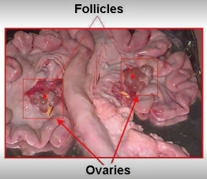
The primary structures of the female reproductive tract are the ovaries, they have two major functions:
1. Produce ova, the female germ cells and
2. Produce the hormones progesterone and estrogen.
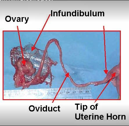
Each ovary is surrounded by a thin membrane called the infundibulum, which acts as a funnel to collect ova and divert them to the oviduct. The oviduct is about 6-10 inches long and acts as the site of fertilization.
– Photos courtesy of Purdue University
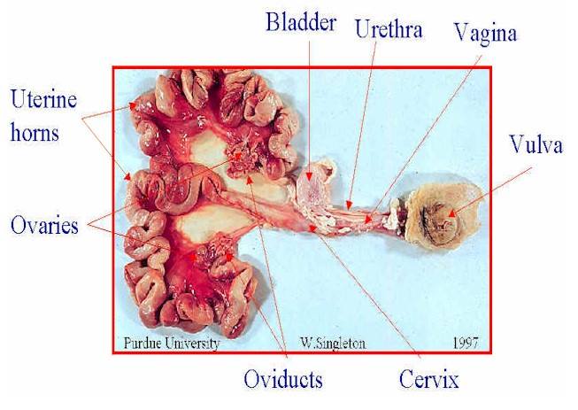
There are two uterine horns. Each is 2-3 feet in length in the non-pregnant sow. They act as a passageway for sperm to reach the oviduct and are the site of fetal development. The uterine body, which is small compared to some other species, is located at the junction of the two uterine horns.
The cervix is a muscular junction between the vagina and uteri. It is the site of semen deposition during natural mating and AI. It is dilated during heat (estrus) but constricted during the remainder of the estrous cycle and during pregnancy.
The vagina extends from the cervix to the vulva and serves as a passageway for urine and the piglets at birth. The bladder is connected to the vagina by the urethra.
The vulva is the external portion of the reproductive tract. It often becomes red and swollen just prior to estrus and this swollen condition is usually more pronounced in gilts than in sows.
The hypothalamus located at the base of the brain secretes gonadotropin releasing hormone (GnRH) which regulates the anterior pituitary gland to secret FSH (Follicle Stimulating Hormone) and LH (Luteinizing Hormone) into the blood which stimulates the production of the ovarian hormones, estrogen and progesterone, which in turn regulate the reproductive process. Oxytocin is released from the posterior pituitary gland.

– photo courtesy Purdue University


