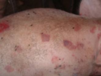
Not-In-Pig (NIP)

We all get frustrated when our sow/gilt return and we find that they are not in-pig (pregnant). The cause of this is not being vigilant in watching if they return (come back on heat, hogging) or it could be a case of an infection.

Causes

The main causes are poor management and infertility. With regards to management, experience shows us that empty sows and gilts are as of a result of being misidentified poorly, housed in groups running with the boar and services are not supervised and observation of post service for returns to oestrus are not detected. I know it is difficult to keep an eye on our pigs all the time, but if you know when they are due to start hogging then introducing the boar and observing can save you time and worry. Knowing if and when the sow/gilt is due to return by counting 21 days from her last hogging will help you to know if the service has been successful.

If pregnancy testing is not carried out (hire our pregnancy scanner from our website oxfordsandyblackpiggroup.org), then sows may reach term without being pregnant. Infertility may be solely responsible when observation has been a little lax. When embryos are produced and die, sows return to oestrus at uneven intervals. Where disease or cold have reduced the ability of the animal to return or where housing is such that hogging cannot be observed, then the pigs will remain undetected until a routine pregnancy check is followed or the expected farrowing date arrives with no farrowing. Sometimes, in these cases, there may be an undetected abortion, cystic ovaries or pyometra which there will be an infection of the uterine horn.

Clinical signs

‘Not-in-pig’ sows or gilts return to heat after service and also fail to develop the abdominal swelling and underline development typical of pregnancy. The sows are found to be non-pregnant when examined by. A history of vulva discharge may pinpoint the stage at which pregnancy ended.

It will come to no surprise that failure to farrow leads to a diagnosis of not-in-pig. The reasons for the failure of pregnancy can be determined by examining the records. If the recording is not carried out, services are not supervised, pregnancy checks not carried out and returns to oestrus not checked visually, then poor management is the cause.

Pigs that are in poor condition and exposed to too hot, cold, damp and drafty housing then this will result in the sow/gilt not carrying to term and possibly reabsorbing or aborting. Where records confirm that service was carried out and pregnancy has been confirmed reliably, then again, abortion or reabsorption has occurred.

Treatment and prevention

Before animals are treated as not-in-pig, a pregnancy test should be carried out, as the commonest cause of failure to return to service is pregnancy. Cystic ovaries and subdued oestrus can be treated using chorionic gonadotrophin. Pyometra may respond to antimicrobial treatment by injection and allow a return to oestrus.

Prevention is largely a matter of management, although vaccination against conditions such as parvovirus, erysipelas, leptospirosis, PRRS, and influenza can also be a reason for a non-productive sow.

Sows must be identified individually, services must be attended with good recording keeping of the boar or semen used together with results of any pregnancy tests. Ensure that mating is successful and occurs at the correct time if AI-ing that semen quality is adequate. Be vigilant to observe that there are no vulva discharges after service.

To hire our pregnancy scanner, please click here

Mastitis

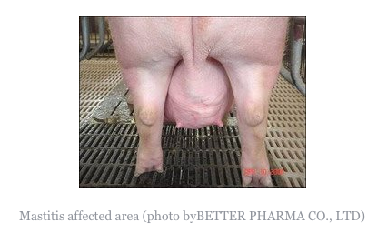
Most of us have experienced this in our sows and it also affects the gilts. There are a few causes to mastitis such as teat injuries from floors or piglets, bacterial infection, poor hygiene and the symptoms is that the udder is hot, hard and swollen, lumps and sometimes the sow/gilt may become unwell and the piglets may also suffer due to lack of feeding.

Causes

Mastitis in sows is caused by ascending infection of the teats by bacteria. The organism most commonly involved is Escherichia coli or related organisms (coliforms). Bacteria such as Arcanobacterium pyogenes, streptococci and staphylococci may cause infections of single glands. Acute and severe mastitis caused by Klebsiella spp. may occur in outbreaks of fatal disease following trauma to the teats caused by rough sawdust bedding. Traumatic injury resulting from piglet teeth, sawdust bedding, or poor quality flooring add to infection. One or both glands supplying a single teat may be infected. Infection enters via the teat canal following teat contamination and bacteria multiply in the gland. Introduction of endotoxin into the mammary gland will cause mastitis and agalactia and endotoxin can be detected in the blood in mastitis.

Clinical signs

Acutely affected sows are usually depressed, loss or lack of appetite and fevered (temperatures of 40.5-42°C, 105-107°F are not uncommon). The udder is usually swollen and oedematous (abnormal swelling of fluid), often with massive congestion. Any secretion that may be obtained after oxytocin injection contains pus. Pain in the udder may lead to restlessness in the sow when piglets attempt to suck. The litter rapidly loses condition. Acute mastitis usually occurs within 1-3 days of parturition. The body temperature falls below normal, the animal can no longer rise and respiratory distress develops frequently leading to death. Coliform (a bacteria calledaerobic bacillus that lives in the colon) mastitis appears to regress within 3-4 days although in severe cases lactation may cease entirely. Sub-acute infection or infection in one or more glands occurs much more commonly and may be recognised by the increased hardness of the gland and, in its early stages, by a square area of reddening or the skin over the affected gland. The litter loses condition. Mastitis in a single gland is often noted only when an affected gland fails to return to normal after weaning. There is often teat injury, especially in the pairs of teats.

Acute mastitis is easily recognisable as affected sows are off their feed and have obvious swelling of the udder, sometimes with reddening and oedema. The litter is usually in poor condition. Animals are often lying down alot, but must be made to rise in order to confirm that mastitis is present. The udder should be felt on both sides by running the hand under both lines of glands.

Individual affected glands feel firm and hot. Laboratory examination of expressed secretion confirms that presence of mastitis by means of cell counts and the organism(s) responsible can be identified. Action requires oxytocin injection as milk let down is under voluntary control. Sub-clinical mastitis may only be detected after cell counts on expressed milk, showing 75% white blood cells. Chronic mastitis is easily felt upon inspection during lactation and easily seen in dry sows.

Treatment and prevention

Acutely ill sows may be saved by injection with neomycin, tetracyclines, ampicillin, amoxicillin, streptomycin, fluoroquinolone, ceftiofur or trimethoprim: sulphonamide for 2-4 days. Oxytocin should also be given. The litter should be fostered or reared artificially. After recovery it may be necessary to cull the sow as she will be prone to this ailment on all farrowings.

Other supportive treatment may include anti-inflammatory medication. In sub-acute cases, rehydration is not necessary. Antimicrobial injection may improve the condition where single glands are affected, but results are poor where abscess are present and has occurred or teat ends are damaged.

Control depends upon hygiene, use of soft bedding other than sawdust, early treatment and, possibly, the use of commercial E. coli vaccine if the condition is due to E. coli. Where the condition is recurring and pre-farrowing infection is suspected, treatment with trimethoprim sulphonamide at 15 mg/kg given in the feed from day 112 of gestation to day 1 post-partum may eliminate early lesions and prevent the development of clinical disease. Sows which have had severe or repeated bouts of mastitis should, unfortunately, be culled and affected sows should only be retained if they have sufficient functioning teats.

Meningitis

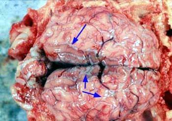
The brain and spinal cord are protected within bony cavities (the skull and the spinal column) but separating the nervous tissue from the bone is a series of membranes called the meninges. When these become inflamed in the condition known as meningitis (usually due to bacterial infection), pressure builds up on the nervous tissue and nervous signs ensue.

In the early stages of meningitis, pigs will be dull and depressed, reluctant to stand and have a raised rectal temperature. Occasionally, they may be seen pressing their head against a wall and they will be unsteady on their legs. As the disease progresses and in response to stimulation (handling, noise), affected pigs will subside into paddling convulsions while lying on their side. Careful observation will reveal that the eyes, when open, will flick from side to side (this is called nystagmus). Death can ensue within a few hours and in some cases of meningitis may simply be found dead.

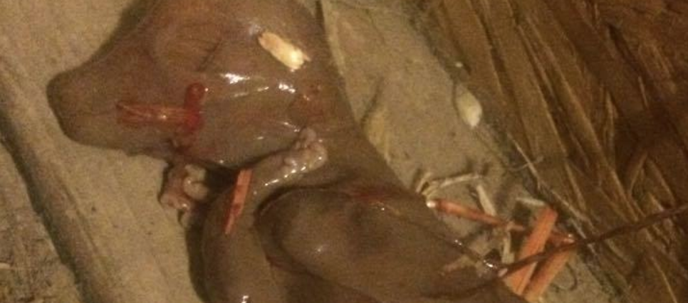
Infection in the brain results from the bloodborne spread of bacteria, which can gain entry through any break in the skin or mucosa. Sporadic meningitis is particularly seen in the young piglet, where infection gains entry through the navel, tail-dock wound, clipped teeth or fight wounds, and is very much a feature of colostral insufficiency. Bacterial spread in the bloodstream (bacteriaemia) can lead to infection in other areas of the body – particularly the joints, causing joint ill, or arthritis. The bacteria involved are usually environmental contaminants such as staphylococci, E. coli and streptococci. However, epizootic forms of meningitis can occur, particularly in weaner pigs four to ten weeks old and rarely in older growing pigs. The most common cause in these cases is Streptococcus suis type II and Haemophilus parasuis.

Streptococcus suis Meningitis

This is typically a post weaning disease that is triggered by the stress of weaning and by mixing pigs of different ages. Overcrowding, poor ventilation and in particular, high humidity all seem to exacerbate the disease. In an infected herd, the organism is picked up at or soon after birth, the reservoir being the nasal chambers and tonsils as well as the vagina of the sow. The organism colonises the tonsil of the young pig and from there will spread via the bloodstream to the brain.

Onset of the disease can be extremely rapid and sudden death may be seen. Streptococcus suis can readily be cultured from the meninges of an affected pig that has not been treated. Paddling convulsions are a classic feature, along with temperatures of 41degrees C (106f) and above. The disease usually behaves in an all or nothing way – in contrast with most gut or respiratory tract diseases, there is no effect on the growth of unaffected meningitic pigs.

Control can be difficult, vaccines have not proved effective and strategic use of antibiotics such as amoxycillin by injection at weaning may be needed to prevent clinical disease. Medication of weaned pigs via water or feed with penicillin-based antibiotic will often suffice in milder outbreaks. Recovered pigs may drop dead suddenly two or three weeks later, the result of seeding of infection on the heart valves and production of endocarditis.

Worth noting that S. suis type II (along with other strains of S. suis) are zoonotic, so particular care should be taken when handling affected pigs and when attending farrowing in herds known to be infected. Infection for humans is normally by skin penetration through cuts and grazes – thorough hand washing with soap is essential following contact with potentially infect material.

Haemophilus parasuis Meningitis

There are many strains of H. parasuis present within pig populations and, while the most common manifestations of disease are either respiratory or septicaemic, cases do occur where the infection targets the brain, producing meningitis (arthritis is also occasionally seen). The clinical presentation is identical to that described above, but diagnosis can be difficult as the organism is very fragile post mortem – euthanasia of an affected untreated pig and immediate sapling of the meninges or cerebral spinal fluid is essential.

Treatment

Treatment of any form of meningitis is based upon killing the causative organism and providing support therapy. Streptococcus suis is generally very sensitive to antimicrobials treatment with penicillin-based medicines such as amoxycillin, but treatment must be rapid and involve a formulation that achieves high levels of antibiotic in the body immediately. While Haemophilus parasuis is also very sensitive to antibiotics, response to treatment of affected pigs is frequently disappointing, probably due to the fact that clinical signs are the result of toxin release rather than the effect of the bacterium itself; by the time signs are seen, the damage is done and killing off the organism will have no effect.

A common complication of meningitis in pigs is dehydration. Their inability to feed and drink soon leads to fluid shortage, especially in the young pigs. This is often manifests in the form of “salt poisoning” (water-deprivation neuropathy), which can easily be confused with meningitis clinically even though it is quite distinct from it. So many pigs that die through meningitis infection actually subside into salt poisoning, which is the true cause of death.

The pig should be removed from the rest of the litter – where it can be bullied – and placed in a bedded area to prevent injury. Often, response to treatment will be very rapid (within two to four hours). It should be given fluids (water containing electrolytes), preferably by mouth on a little and often basis. Care must be taken to ensure that the pig is swallowing fluids and not inhaling them.

Support treatment with corticosteroids or non-steroidal anti-inflammatory agents can also improve recovery rates. As a general rule, however, a failure to respond to treatment within forty-eight hours is likely to leave the pig permanently brain damaged and so euthanasia is appropriate.

IN ALL CASES PLEASE DISCUSS YOUR SITUATION WITH YOUR VETERINARY PRACTICE.

Photos obtained from Google

Porcine Parvovirus (PPV)

PLEASE REMEMBER TO SIGN UP TO THE DISEASE CHARTER WITH AHDB PORK.

When sourcing stock it is up to you to ask the breeder if they have suffered any diseases within their herd if you don’t ask you don’t know and the breeder will and should not be offended by your question as this shows due diligence on your behalf.

It it my intention to discuss diseases specific to the pregnant sow and the effects her infection will have on the unborn litter and then move on to respiratory disease, nervous system disease etc over the weeks. So hope you find it all helpful.

Reproduction Disease – Porcine Parvovirus (PPV)

Apart from a single very unusual report of skin disease in weaners, PPV is solely associated with the reproductive failure or with its effect on an unborn litter. Infection of the non-pregnant animal has no clinical effect and immunity is acquired that will be life-long and will protect all future litters. The effects that PPV infection will have on a pregnant sow depend on the stage of pregnancy, which I have shown below.

Stage of reproductive cycle Effect of PPV infection Result

Not pregnant No effect Immunity

At service and within Death of fertilised eggs/ Return to service 10 days of service differentiating embryos at 3 weeks 10 – 25 days post service Embryonic death Delayed return to service or small litter 25 – 75 days post-service Foetal death, often Variable-sized mummified progressive through the litter pigs affecting whole or part of the litter, and/or stillbornpigs

75 days plus Minimum effect on foetuses Possibly small pigs born as the immune response can be that have been checked during growth, and stillborn pigs

From this it can be seen that the effects of PPV infection can be stillbirth, mummification, embryonic death and infertility, giving the old acronym SMEDI. It should be noted that abortion is a very rare manifestation of PPV infection.

On a herd basis, in a naïve herd, an outbreak of PPV disease will last two to three months and will manifest by varying signs over that time in the following sequence:

  1. Increased regular returns to oestrus lasting two to three weeks
  2. Increased irregular returns to oestrus lasting two to three weeks simultaneously with above
  3. Stillborn pigs starting simultaneously with above for one to two months.
  4. Increase in mummified pigs from six to twelve weeks after (1) above, and failures to farrow.
  5. Drop in total litter size for two to three weeks from eighty days after the start of the outbreak.

It is a sequential disease, the classic sign of which is large numbers of mummified pigs within a litter, of variable size, starting around one month after an increase in returns to service. The diagnosis of PPV disease is based on clinical pictures supported by blood tests and by virus detection in the livers of mummified or stillborn pigs.

Prevention and Control

Highly effective vaccines against PPV, which we have discussed in the previous post on the group, are available and are given to gilts prior to breeding. The actual programme (number of doses and timing) varies between products, but it should be noted that maternally derived antibodies (those passed to a piglet in colostrums) can survive for up to six months and that these can block vaccine efficacy. Vaccination should, therefore, not be given too early in life. It is also worth noting that the disease can be transmitted from boars to sows or vice versa by direct mating with infected pigs or through artificial insemination from infected semen of boars.