PPV, while not outwardly noticeable in our pigs during day-to-day observation, poses significant challenges as it affects our in-pig sows and gilts. This virus can wreak havoc internally, impacting embryo and foetus development.
Previously known as “stillbirth mummification embryonic death and infertility” (SMEDI), PPV was once a prevalent issue. However, effective vaccines have considerably reduced its occurrence.
Signs and Effects:
Signs of PPV vary depending on the stage of pregnancy. Infection can occur during mating, leading to total embryo loss and subsequent return to estrus. Later infections may still result in embryo destruction, with pregnancies terminating if fewer than four embryos are present. As the virus progresses, it can lead to termination of all foetuses, initiating a process of re-absorption and resulting in mummified births.
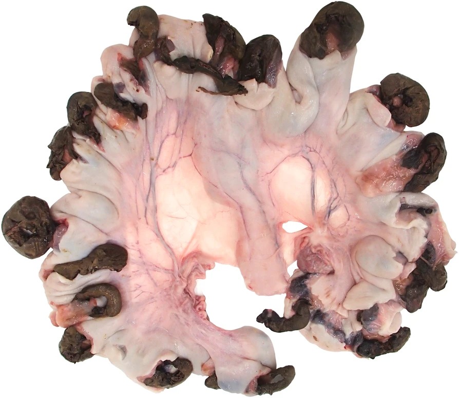
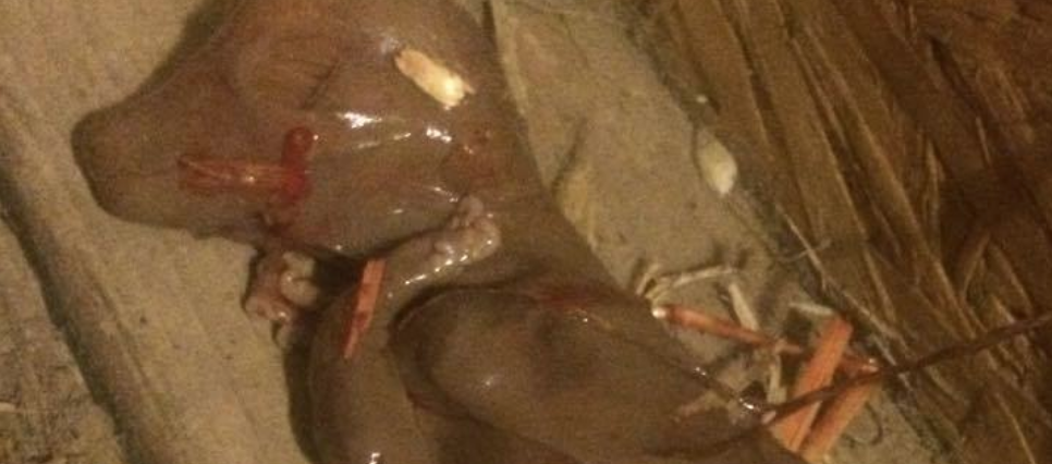
This litter belonged to a non-vaccinated gilt that was confirmed with PPV at approximately day 40 of gestation. Photo: Boehringer Ingelheim Vetmedica Inc.
Diagnosis and Behaviour:
Diagnosing PPV involves observing certain behaviours in breeding sows and gilts, such as repeated hogging every 3 weeks or irregular patterns every 4-5 weeks, along with the presence of mummified piglets and stillbirths.
Prevention and Vaccination:
Preventing PPV primarily relies on a robust vaccination program. Vaccines, particularly the combi vaccine Ery/Parvo, are highly effective. Consulting with your veterinary practice is essential for guidance on vaccination protocols, including vaccinating gilts prior to service and providing booster doses as recommended.
Considerations for Boars:
Boars themselves may not show clinical signs or semen damage from PPV but can carry and transmit the virus, potentially infecting gilts. Some veterinarians recommend vaccinating boars every six months to mitigate this risk, particularly in AI studs.
For comprehensive prevention and control strategies against PPV, veterinary advice and guidance is paramount.
The Oxford Sandy and Black Pig Group is UK’s only pig breed that is a registered charity in England & Wales (1190469) and Scotland (SCO52662). We are creating a better future for our breed the bloodlines and it breeding potential together with our Independent Pork Producers, Breeders and Oxford Sandy and Black Keepers and their families. Please click the donatebutton so we may continue to look after our breed and our supporters
Follow us on Facebook and see how we support, help and inspire individuals about our rare breed
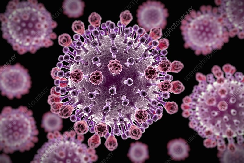
Similar to humans, pigs are susceptible to colds and flu, especially in the Autumn and Winter seasons; however, they can contract these illnesses at other times of the year as well. In pigs, this condition is referred to as Swine Influenza (swine flu), characterised as a respiratory disease caused by type A influenza virus. Swine Influenza regularly sparks outbreaks in pig populations. While swine flu viruses typically do not infect humans, rare cases of human infections have been reported. In pig herds, these viruses can lead to significant illness, although fatalities are infrequent.
The virus circulates among pigs throughout the year, with a tendency to peak during late Autumn and Winter months, mirroring the seasonal patterns observed in human flu and colds.
Similar to influenza viruses in humans and other animals, swine flu viruses undergo frequent changes. Pigs have the susceptibility to be infected by avian flu and human flu viruses. When flu viruses from diverse species infect pigs, mutations can occur, giving rise to new viruses that are a combination of swine, human, and/or avian flu viruses. This continual evolution has led to the emergence of various variations of swine flu viruses over the years. As a result, there are three primary influenza A virus subtypes that have been identified in pigs: H1N1, H1N2, and H3N2.
The primary mode of transmission for swine flu viruses among pigs is believed to be close contact, and there is a potential for transmission through contaminated objects moving between infected and uninfected pigs. Swine herds, even those that have been vaccinated against swine flu, may experience sporadic disease, with some showing only mild or no symptoms of infection.
Symptoms Clinical manifestations of swine flu in pigs may encompass symptoms such as fever, depression, coughing (barking), discharge from the nose or eyes, sneezing, breathing difficulties, eye redness or inflammation, and a decrease in appetite. It is noteworthy that certain pigs infected may not display any signs of illness.
Is Swine Flu common
Before 1985, Swine Flu had not clinically appeared in pigs in the UK. However, subsequent investigations revealed the presence of a specific strain, H3N2, derived from humans, in the pig population since 1968. This strain was first associated with clinical disease in pigs in 1986, a year after cases of Swine Flu caused by a pig-specific H1N1 strain emerged. Later, in 1991/2, a new strain emerged, a mutation of the H1N1 pig strain and an avian influenza strain, designated H1 A/SW/195852/92. This strain, combined with PRRS (Blue Ear) infection, proved particularly severe. Throughout the 1990s, additional strains such as H1N2 and H3N1 were detected in pigs.
At present, the disease is widespread throughout the UK, manifesting as sporadic cases in individual herds, occasionally across regions, and persistently affecting certain farms for several months. Reports from APHA covering the winter period of 2015/6 indicate common disease outbreaks, primarily attributed to either the human pandemic strain A H1N1 09v or H1N2. Similar to human influenza, its prevalence peaks during winter conditions when virus survival rates are high, although cases can occur at any time of the year. A significant incidence of the pandemic strain in humans heightens the risk of widespread infection among pigs.
Treatment
Treatment for influenza in pigs is limited due to its viral nature, and unless there is a secondary infection, treatment is generally unnecessary. Typically, pigs will recover within a week. However, supportive therapy using aspirin administered via the water system or paracetamol in feed may aid in accelerating individual pig recovery. (As stated from NADIS) However in all cases do seek advice from your vets.
Nevertheless, Swine Flu, particularly in grower pigs, can make them vulnerable to other diseases, notably Actinobacillus pleuropneumoniae. Many outbreaks of the latter have been linked to preceding unrecognised Swine Flu outbreaks. In such instances, prompt treatment with appropriate antibiotics is crucial to mitigate potentially catastrophic losses associated with this disease.
During uncomplicated Swine Flu outbreaks, growth may be slowed, and affected pigs may not be suitable for slaughter while they are ill. It is important to avoid overcrowding during outbreaks, and slaughter weights may be reduced after the disease has subsided.
Worth Noting: For over two decades, Swine Flu has posed a significant concern to pig keepers in the UK, with varying strains causing a range from mild to severe, including loss of appetite and respiratory issues. The emergence of the more recent human pandemic strain has heightened concerns among pig keepers due to its potential as a reverse zoonosis. This strain not only presents a direct threat as a primary source of disease but also raises alarms regarding its ability to intermingle with existing flu strains in pigs. Such interactions could potentially give rise to even more potent strains, posing risks to both pig and human populations alike.
Attention to high standards of biosecurity, isolation of incoming stock away from your main area of your holding/Farm have parts to play in the prevention of many diseases. Not allowing to share same air space and no nose-to-nose contact. Biosecurity signs obtainable HERE from AHDB
The Oxford Sandy and Black Pig Group is UK’s only pig breed that is a registered charity in England & Wales (1190469) and Scotland (SCO52662). We are creating a better future for our breed the bloodlines and it breeding potential together with our Independent Pork Producers, Breeders and Oxford Sandy and Black Keepers. Please click the donatebutton so we may continue to look after our breed and our supporters
Follow us on Facebook and see how we support, help and inspire individuals about our rare breed
We all get frustrated when our sow/gilt return and we find that they are not in-pig (pregnant). The cause of this is not being vigilant in watching if they return (come back on heat, hogging) or it could be a case of an infection.
Causes
The main causes are poor management and infertility. With regards to management, experience shows us that empty sows and gilts are as of a result of being misidentified poorly, housed in groups running with the boar and services are not supervised and observation of post service for returns to oestrus are not detected. I know it is difficult to keep an eye on our pigs all the time, but if you know when they are due to start hogging then introducing the boar and observing can save you time and worry. Knowing if and when the sow/gilt is due to return by counting 21 days from her last hogging will help you to know if the service has been successful.
If pregnancy testing is not carried out (hire our pregnancy scanner from our website oxfordsandyblackpiggroup.org), then sows may reach term without being pregnant. Infertility may be solely responsible when observation has been a little lax. When embryos are produced and die, sows return to oestrus at uneven intervals. Where disease or cold have reduced the ability of the animal to return or where housing is such that hogging cannot be observed, then the pigs will remain undetected until a routine pregnancy check is followed or the expected farrowing date arrives with no farrowing. Sometimes, in these cases, there may be an undetected abortion, cystic ovaries or pyometra which there will be an infection of the uterine horn.
Clinical signs
‘Not-in-pig’ sows or gilts return to heat after service and also fail to develop the abdominal swelling and underline development typical of pregnancy. The sows are found to be non-pregnant when examined by. A history of vulva discharge may pinpoint the stage at which pregnancy ended.
It will come to no surprise that failure to farrow leads to a diagnosis of not-in-pig. The reasons for the failure of pregnancy can be determined by examining the records. If the recording is not carried out, services are not supervised, pregnancy checks not carried out and returns to oestrus not checked visually, then poor management is the cause.
Pigs that are in poor condition and exposed to too hot, cold, damp and drafty housing then this will result in the sow/gilt not carrying to term and possibly reabsorbing or aborting. Where records confirm that service was carried out and pregnancy has been confirmed reliably, then again, abortion or reabsorption has occurred.
Treatment and prevention
Before animals are treated as not-in-pig, a pregnancy test should be carried out, as the commonest cause of failure to return to service is pregnancy. Cystic ovaries and subdued oestrus can be treated using chorionic gonadotrophin. Pyometra may respond to antimicrobial treatment by injection and allow a return to oestrus.
Prevention is largely a matter of management, although vaccination against conditions such as parvovirus, erysipelas, leptospirosis, PRRS, and influenza can also be a reason for a non-productive sow.
Sows must be identified individually, services must be attended with good recording keeping of the boar or semen used together with results of any pregnancy tests. Ensure that mating is successful and occurs at the correct time if AI-ing that semen quality is adequate. Be vigilant to observe that there are no vulva discharges after service.
PLEASE REMEMBER TO SIGN UP TO THE DISEASE CHARTER WITH AHDB PORK.
When sourcing stock it is up to you to ask the breeder if they have suffered any diseases within their herd if you don’t ask you don’t know and the breeder will and should not be offended by your question as this shows due diligence on your behalf.
It it my intention to discuss diseases specific to the pregnant sow and the effects her infection will have on the unborn litter and then move on to respiratory disease, nervous system disease etc over the weeks. So hope you find it all helpful.
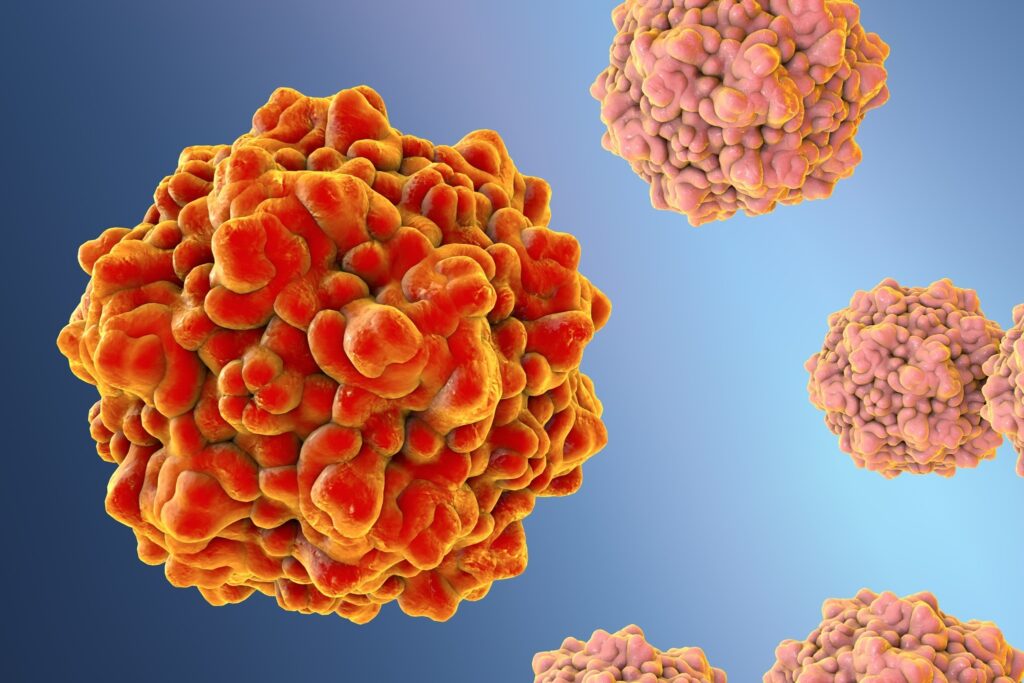
Reproduction Disease – Porcine Parvovirus (PPV)
Apart from a single very unusual report of skin disease in weaners, PPV is solely associated with the reproductive failure or with its effect on an unborn litter. Infection of the non-pregnant animal has no clinical effect and immunity is acquired that will be life-long and will protect all future litters. The effects that PPV infection will have on a pregnant sow depend on the stage of pregnancy, which I have shown below.
Stage of reproductive cycle Effect of PPV infection Result
Not pregnant No effect Immunity
At service and within Death of fertilised eggs/ Return to service 10 days of service differentiating embryos at 3 weeks 10 – 25 days post service Embryonic death Delayed return to service or small litter 25 – 75 days post-service Foetal death, often Variable-sized mummified progressive through the litter pigs affecting whole or part of the litter, and/or stillbornpigs
75 days plus Minimum effect on foetuses Possibly small pigs born as the immune response can be that have been checked during growth, and stillborn pigs
From this it can be seen that the effects of PPV infection can be stillbirth, mummification, embryonic death and infertility, giving the old acronym SMEDI. It should be noted that abortion is a very rare manifestation of PPV infection.
On a herd basis, in a naïve herd, an outbreak of PPV disease will last two to three months and will manifest by varying signs over that time in the following sequence:
Increased regular returns to oestrus lasting two to three weeks
Increased irregular returns to oestrus lasting two to three weeks simultaneously with above
Stillborn pigs starting simultaneously with above for one to two months.
Increase in mummified pigs from six to twelve weeks after (1) above, and failures to farrow.
Drop in total litter size for two to three weeks from eighty days after the start of the outbreak.
It is a sequential disease, the classic sign of which is large numbers of mummified pigs within a litter, of variable size, starting around one month after an increase in returns to service. The diagnosis of PPV disease is based on clinical pictures supported by blood tests and by virus detection in the livers of mummified or stillborn pigs.
Prevention and Control
Highly effective vaccines against PPV, which we have discussed in the previous post on the group, are available and are given to gilts prior to breeding. The actual programme (number of doses and timing) varies between products, but it should be noted that maternally derived antibodies (those passed to a piglet in colostrums) can survive for up to six months and that these can block vaccine efficacy. Vaccination should, therefore, not be given too early in life. It is also worth noting that the disease can be transmitted from boars to sows or vice versa by direct mating with infected pigs or through artificial insemination from infected semen of boars.
When dealing with large litters and you have simultaneous farrowings, there may be an opportunity to implement a fostering procedure, transferring your Oxford Sandy and Black piglets from a larger farrowing to a smaller farrowing. This practice can be of benefit as well as ensuring the welfare of sow and piglets which begins with careful observation of the piglets.
Identify those piglets that show signs of lethargy and lacklustre. These observations will serve as indicators for taking action, possibly including the decision to foster the struggling piglet and at the same time provide essential sustenance such as electrolytes or additional milk.
Cross-fostering your low birth weight Oxford Sandy and Black piglets with littermates of similar weight can significantly improve their daily weight and overall health with the added bonus of high expectations for pre-weaning performance.
We are aware that smaller piglets competing with their larger siblings are at a disadvantage. Therefore, whenever possible, it is advisable to create foster litters consisting of the smallest piglets born on a given farrowing day. It is essential to introduce these small piglets to a sow that is in her early stages of motherhood, preferably on her first or second litter. Sows in their second year, for instance, tend to have smaller teats that are better suited for the small mouths of these piglets. This thoughtful approach can greatly benefit the piglets’ growth and overall well-being of the Oxford Sandy and Black Pig.
You might discover our blog titled “Piglet/Weaner – Being Prepared” to be a valuable resource for additional guidance and assistance.
The Oxford Sandy and Black Pig Group is UK’s only pig breed that is a registered charity in England & Wales (1190469) and Scotland (SCO52662). We are creating a better future for our breed, the bloodlines and its breeding potential together with our Independent Pork Producers, Breeders and Keepers. Please consider clicking our donate button so we may continue to look after our breed and our supporters.
Follow us on Facebook and see how we support, help and inspire individuals about our rare breed
Photo: In pig gilt with 11 days to go. Francesca an Oxford Sandy and Black Pig Gilt photo taken on 22 January 2023 farrowed on 2 February 2023.
Enhancing the In-pig Sow Nutrition
Although the last month of pregnancy is the period when the main growth rate of the foetus takes shape, we, Oxford Sandy and Black Pig keepers must take into consideration the whole of the gestation period to take advantage of the opportunity to influence birth weights.
Direct Your Attention Beyond the Final Third of Gestation: Emphasis is more than just the last third of gestation, as this period witnesses foetal growth spurts and an opportunity to influence birth weight. Elevate nutrient intake for sows starting from day 80 of gestation to positively impact birth weight. Notably, between days 90 and 115, piglet growth rate increases significantly.
Evaluate Feeding Regime for the Oxford Sandy and Black Gilts: Carefully consider the feeding plan for your breeding gilts. Overfeeding them during the initial stages of gestation can yield both short and long-term effects. The extra feed might be directed towards the gilts’ reserves rather than the developing foetus. This practice could lead to the birth of small piglets and overweight gilts. Once a gilt accumulates excess fat, her productivity might diminish.
Remember it is worth discussing feeding options with your feed nutritionist/feed merchant.
Photo: In pig gilt with 12 days to go. Francesca an Oxford Sandy and Black Pig Gilt photo taken on 22 January 2023
The Oxford Sandy and Black Pig Group is UK’s only pig breed that is a registered charity in England & Wales (1190463) and Scotland (SCO52662). We are creating a better future for our breed, the bloodlines and its breeding potential together with our Independent Pork Producers, Breeders and Keepers. Please consider clicking our donate button so we may continue to look after our breed and our supporters.
Follow us on Facebook and see how we support, help and inspire individuals about our rare breed
As we witness the wonderful farrowings with the anticipation of more to come, I thought it a perfect opportunity to delve into the different phases of piglet observation and care this week.
We know that recognising those piglets smaller than their littermates often exhibit delayed growth and require additional attention, and as such the following protocols may aid us to minimise the weight disparity between lighter piglets and their larger siblings.
Investigation of various factors affecting birth weight, with the aim of enhancing the postnatal performance of underweight piglets, has resulted in the following findings.
As most of us have witnessed, a common occurrence is litters with diverse birth weights. These variations tend to increase, with an overall trend toward smaller piglets, when the number born alive is higher. There is no need for alarm; entire litters comprised solely of small piglets do not necessarily indicate an issue, as these piglets can still be viable. With the right conditions, low birth weight pigs can catch up in growth to their normal birth weight counterparts. Nonetheless, early intervention remains crucial.
The prospect of piglet survival rates are low if their weight falls below 1 kg. If possible, try to weigh and gauge the number of piglets below 1kg, document the weights to enable to recognise the developments which will help you to offer extra assistance in their development.
Challenges During Foetal Development
Poor foetus and reduced growth of piglets can be identified as early as 30 days of gestation
Low birth weight can be a result of an inefficiency of the placenta to transfer nutrients to the foetuses rather than uterine capacity – ensure gilt is not younger than 12 months for service
Oxygen Deprivation influences the Critical Factor in Foetal Growth, Central Nervous System Impact, and Survival of Piglets
Some piglets may be petite but exhibit excellent vitality and favourable postnatal behaviours, such as rapid suckling, which significantly contributes to their survival.
Piglets that are both undersized and weak, which can be indicative of oxygen deprivation, face a significant disadvantage
It’s important to highlight that the maternal genotype plays a significant role in determining placental efficiency and, consequently, foetal weight, while the potential for growth and size is influenced by the sire line.
The Oxford Sandy and Black Pig Group is UK’s only pig breed that is a registered charity in England & Wales (1190463) and Scotland (SCO52662). We are creating a better future for our breed, the bloodlines and its breeding potential together with our Independent Pork Producers, Breeders and Keepers. Please consider clicking our donate button so we may continue to look after our breed and our supporters.
Follow us on Facebook and see how we support, help and inspire individuals about our rare breed
Some of us have experienced our sows and gilts as being very drawn during and after feeding their piglets. The causes have been diagnosed as inadequate feed during lactation, farrowing house being too hot or is due to underfeeding during pregnancy. This effects weight loss and anoestrus (poor return to season).
Causes
The condition results classically from a combination of parasitism – one organism (called parasite) benefits at the expense of another organism usually of different species (worms or mange), low environmental temperatures and inadequate feed intake, particularly during lactation. Weight loss at this time may never be regained. Parasitism is less important when therapy is routinely carried out. In outdoor pig populations, low environmental temperatures are important.
Outdoor sows require at least 200 kg more feed per sow per year than indoor animals. Sow that are kept indoors during the winter cold months should be kept at 22°C and sows in moderate condition at 21°C. Lower temperatures may be tolerated in the presence of bedding. Temperatures below this require extra feed to maintain sow weight gain in gestation.
Feed intake is the most important cause. Feed intake may be affected by bullying, disease, a high environmental temperature in the farrowing accommodation, or an over fat condition at farrowing. Sows should be in condition score 3/4 or 6/10 (fat depth at P2 16-20 mm) at farrowing, which may fall to condition score 2-2.5/4 or 4/10 (fat depth P2 12 mm) after lactation. Unless these criteria are met, total weaning weight of the litter will be reduced, return to oestrus will be delayed and egg numbers will be reduced to give small subsequent litters.
Clinical signs
In extreme cases, being abnormally thin or weak may occur in 30-90% of sows and boars in a herd, associated with hypothermia 36.5-38°C (97-100°F), depraved appetites, restlessness, apathy, and later, difficulty in rising. The skin may be dirty and greasy and there may be surface abrasions. As the condition progresses, failure to return to oestrus and permanent infertility may occur.
Where accurate individual feeding to condition is not practised and bullying occurs, individual animals in a group can be clinically affected. Less than optimal condition are frequently found in winter where temperature control is not practised. Suboptimal condition or even thin sows may be found during the recovery period from disease such as influenza and may be more extreme in lean breeds. Clinical signs of suboptimal condition include increased weaning to service intervals, small litters and low weaning weights. Piglets of sows in suboptimal condition may be restless and demand milk more frequently.
Thin sows may be identified by observation and systematic condition scoring of the herd. Ultrasound is particularly useful for quantitative measurement as scoring is more difficult in older animals due to their conformation. As sows should gain at least 12.5 kg body weight between parities, regular weighing can identify animals in sub-optimal condition. Pressure sores in sows at weaning also indicate poor condition.
Where gilt condition and nutrition in lactation is inadequate, the second litter is the same size or smaller than the first, and low number born may reflect overall sow condition at service. Extended weaning to service intervals and low weaning weights may be due to poor body condition. The causes of poor sow condition should be established. It should be established that feed of adequate quantity and nutrient density is being supplied to each individual especially during lactation and until implantation 10-14 days after service. Parasitism can be ruled out by inspection, by sampling for mange and faecal sampling (which some of you do) for worm eggs and coccidia. The influence of disease may be established by inspection, clinical examination or consultation of the recent history of the herd of the animals concerned.
Treatment and prevention
There is no treatment. The effects of thin sows on litter weaning weights can be reduced by supplementary feeding of the litter, whereby some of us have supplemented with sow replacement milk in the earlier days. The effects on numbers born may be reduced when sows or gilts are in poor condition by delaying service until the next oestrus. Ensure that adequate quantities of food of the correct nutrient density are given to all affected animals and that individual feeding is possible. Environmental temperatures should be restored to normal and anthelminthic or mange treatment should be considered.
Adequate feed intake during lactation and early pregnancy should maintain the weight of the sow and increase it by 10-15 kg between litters by increasing the energy content of lactating rations with fat or feeding three times daily during lactation. It may be necessary to weigh sows after weaning (for those of you that do not have scales please may I remind you that we have a “calculate the live weight” in the OSBPG tools section of our website) in order to ensure that adequate weight gain has occurred between lactations, to assign them to groups for feeding at an appropriate level through pregnancy or to feed individually to condition.
As we know each breed has its own characteristics. However, a generalisation can be reached regarding selecting breeding stock for future generations. The same criteria applies to the selection of both our boars and gilts/sows.
The ideal pig provides good cushioning and flexability to all the joints and as you would imagine this would cause our pigs an easier time getting up and down with the knock on effect of being less likely to suffer from leg injuries and complaints which in turn enhances the longevity and productivity of our herds regardless of how small or large they will be. Not forgetting adding to a greater genetic selection.
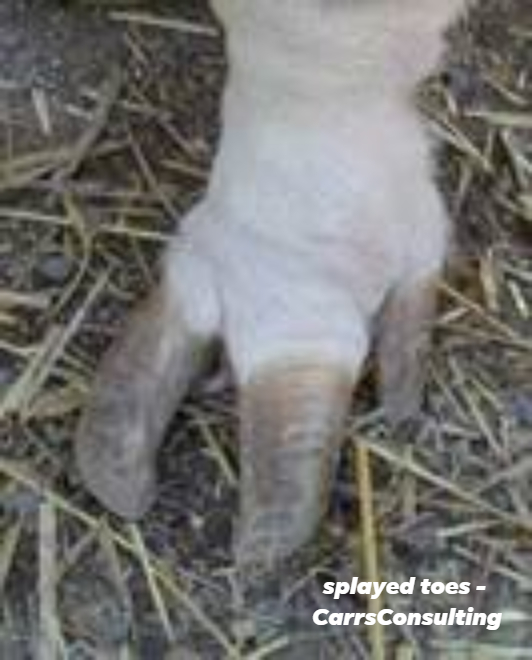
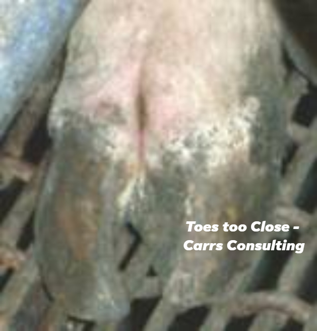
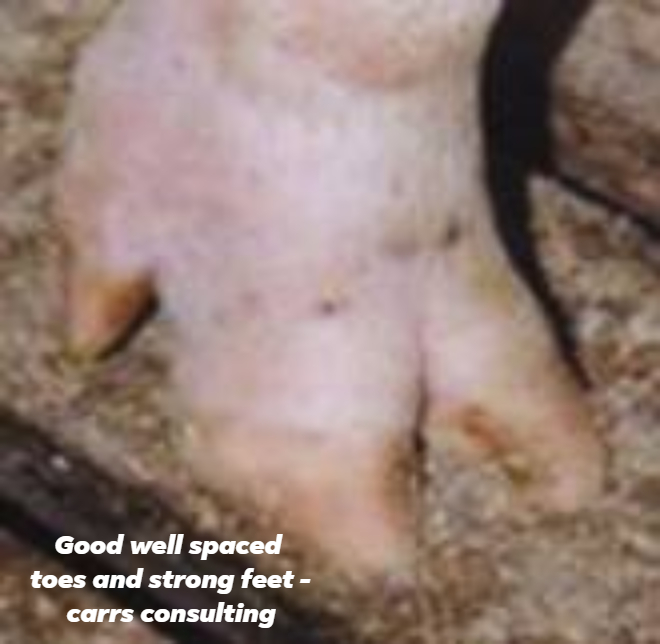
So lets start from the ground up. The toes, the fundamental building blocks of the making of our pigs.
Toes should be big, even and well spaced to take the weight of our pigs.
Condition of the toes
The toes should have no visible cracks, swellings or injuries, this is also true to say for the underneath of each foot.
The Oxford Sandy and Black Pig Group is UK’s only pig breed that is a registered charity in England & Wales (1190463) and Scotland (SCO52662). We are creating a better future for our breed, the bloodlines and its breeding potential together with our Independent Pork Producers, Breeders and Keepers. Please click the donate button so we may continue to look after our breed and our supporters.
Follow us on Facebook and see how we support, help and inspire individuals about our rare breed
Photo: Pork Information Gateway – Faeces with excess blood, mucus or both are highly suggestive of Swine Dysentery
Swine Dysentery, Brachyspira hyodysenteria, a widespread and well-recognised disease among pigs globally, poses a significant threat across various pig keeping operations. It is present in the UK and it is wise that pig keepers are aware of diseases in and around your area/county.
Please ensure you are signed up to AHDB Disease Charter (using the same log in details when signing in for the eaml2). Being aware helps you to be prepared. As it could affect the movement of your pigs.
Clinical Symptoms
Swine dysentery has distinct clinical signs in affected pigs, such as a dull and depressed demeanour, displaying a lack of appetite.
Dehydration is a common consequence of the disease, the faeces may range from soft to almost watery consistency, containing blood, mucous, and, in severe cases there will be necrotic gut lining.
The course of the disease usually spans over several days. Initially, pigs may exhibit high temperatures, reaching up to 41°C (106°F). Mortality can occur early on or later in the disease course, often as a result of dehydration or salt poisoning. It is essential to closely monitor pigs showing these clinical symptoms and promptly seek veterinary attention to manage and treat the disease effectively.
Swine dysentery is primarily spread through infected pigs and their faeces. Anything contaminated with dung, such as vehicles, boots, and equipment, can serve as potential carriers and easily transmit the infection between farms. Vigilant biosecurity measures are crucial in preventing the disease’s spread and protecting pig populations from the risk of infection. Being mindful of where you are moving pigs from and too and being aware of the regional/county infection.
Pigs that survive swine dysentery infection often require treatment, leading to extended time to reach slaughter weight. This compromises the farm’s overall productivity and competitiveness, posing significant challenges for pig producers.
The disease poses a particularly severe threat to farms involved in breeding pigs. If breeding pigs become infected, it can devastate both their international and UK trade, significantly impacting the farm’s economic viability.
An infected pig farm not only endangers other local farms but also poses a risk to farms in other regions. The disease can spread through contaminated vehicles or pig movements, leading to potential outbreaks in previously unaffected areas.
Moreover, the limited range of available treatments for swine dysentery is facing increasing resistance from the disease. In some cases, the swine dysentery organism has become resistant to all available treatments, leaving depopulation as the only viable method to control the disease’s spread and prevent further outbreaks.
The seriousness of swine dysentery demands constant vigilance and the implementation of strict biosecurity measures to safeguard pig populations and the pig farming industry as a whole. Early detection, proper management, and collaboration among pig producers are essential in tackling this challenging and potentially devastating disease.
Prevention
Make use of AHDB free services for safer biosecurity measures and sign up to the Significant Diseases Charter. An important application which shares information quickly in the event of an outbreak.
It is worth noting that abattoirs pose a significant risk for cross-contamination of vehicles. Therefore, effective cleaning and disinfection protocols can successfully prevent such contamination.
If you suspect swine dysentery in your pigs, it is crucial to take immediate action:
Observe for Diarrhoea and Wasting: Pay close attention to any pigs showing signs of unexplained diarrhoea and wasting. In particular, if the diarrhoea contains blood or mucus, this could be an indication of swine dysentery.
Contact Your Veterinarian: As soon as you notice these symptoms, contact your veterinarian immediately for advice and assistance. They will provide guidance on the next steps and help confirm the diagnosis.
Seek Diagnosis: Diagnosis of swine dysentery is achieved by submitting faeces or pig samples for testing. Your veterinarian will conduct appropriate tests to identify the presence of the disease.
Implement Control Measures: If swine dysentery is confirmed, prompt diagnosis is essential for implementing suitable control measures. These measures are critical in limiting the spread of the infection to other pigs and farms.
By taking swift action and involving your veterinarian at the first sign of suspicion, you can effectively manage and control swine dysentery, minimising its impact on your herd and preventing the disease from spreading to other pig populations.
The Oxford Sandy and Black Pig Group is UK’s only pig breed that is a registered charity in England & Wales (1190463) and Scotland (SCO52662) We are creating a better future for our breed, the bloodlines and its breeding potential together with our Independent Pork Producers, Breeders and Keepers. Please click the donate button so we may continue to look after our breed and our supporters.
Follow us on Facebook and see how we support, help and inspire individuals about our rare breed