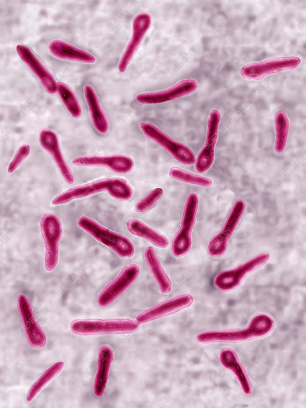
Tetanus is caused by a bacterium called Clostridium tetani, which produces toxins that affect the central nervous system of which the germ lives in the large intestine and can form spores therein. It also exists in faeces, not just in pigs but of other animals, and can also exist in soil.
The incubation period for the disease is around 1 to 10 weeks. Spores of the bacterium can be present in the soil, and the infection usually occurs through a dirty wound or cut. In lactating piglets, castration is a common source of infection.
Young pigs kept outside, especially those with wounds, are more prone to tetanus. Castration wounds in young pigs are particularly at risk. Once infected, the bacterium produces a powerful neurotoxin, leading to rigid muscle contractions.
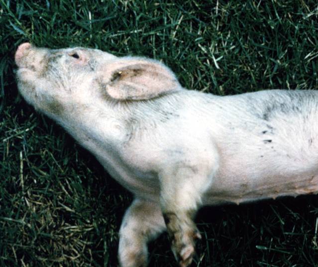
Image: NADIS
Symptoms of tetanus in pigs include collapsing with tetanic muscle spasms, an upright head, curled tips of the ears, all four legs held out backwards, and an upright tail. Death often results from asphyxiation as the respiratory muscles become paralyzed. It is essential not to confuse tetanus with meningitis.
The disease tends to occur in areas known to be infected. In such high-risk situations, it is recommended to remove piglets for 48-72 hours after castration to allow wound closure. Maintaining a clean environment during castration is crucial. The disease is not limited to areas shared with or previously occupied by sheep.
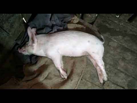
Image: YouTube
In high-risk situations, vaccination of the sow is advisable and should be incorporated into your vaccination program. It is essential to consult with your veterinary for guidance. Consideration of administering a tetanus antitoxin at castration should also be discussed with your veterinary practitioner. Taking these preventive measures can help protect pigs from the potentially deadly effects of tetanus.
Always look to your bio-security. Hygiene is first and foremost.
Visit our website and Become a Friend and support UK’s only pig breed that is a registered charity England & Wales (1190469) Scotland (SCO52662)
We invite you to join us in our mission to preserve and protect the rare breed of Oxford Sandy and Black Pig. Your support is crucial in ensuring a brighter future for this cherished breed, its precious bloodlines, and its valuable breeding potential. Together, let’s work towards providing a thriving and sustainable existence for these remarkable pigs, safeguarding their heritage for generations to come. Your contribution will make a meaningful difference in the conservation and prosperity of the Oxford Sandy and Black Pig breed.
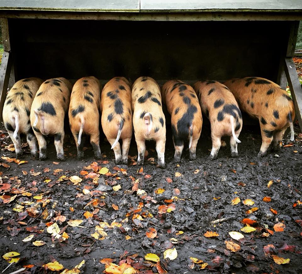
Biting in pigs is not uncommon and can manifest in various forms, such as tail, ear, flank, stifle, anus, belly, vulva, and even penis biting, all of which are different types of cannibalism. However, tail biting is viewed as the most widespread and serious of them all and thankfully rarely seen within the Independent Pork Producer sector. It is true to say that it is most common within the corporate sector. However, it has been observed within the IPP sector, whereby rare/traditional pig breeds are running indoor systems with some experiencing this condition as well a other issues which is underlining the need to seek advice on space and layout of running an indoor system.
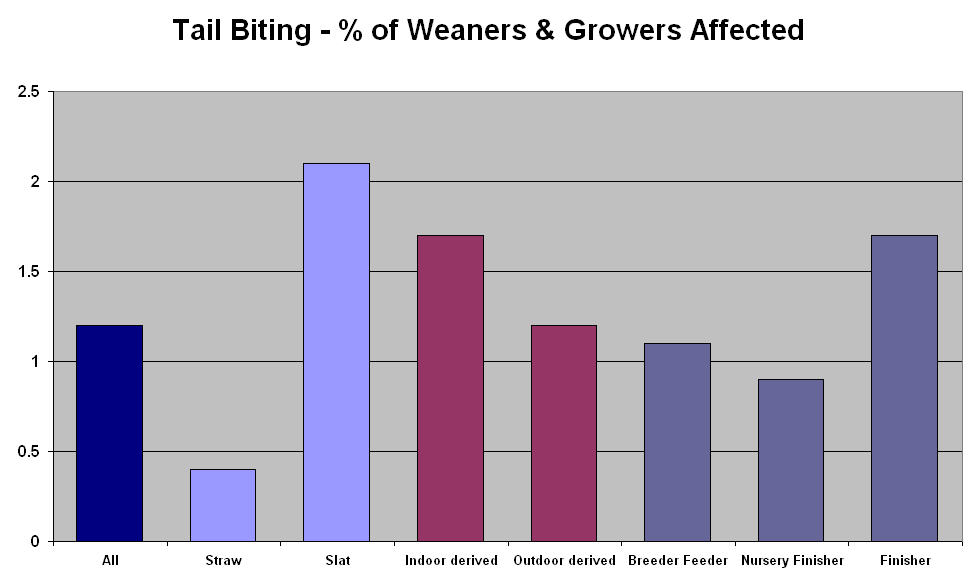
Research has suggested that the overall frequency of tail biting is 1.2% and that the frequency in different systems highlights the widespread nature of the problem. No one system of pig keeping is immune from tail biting. Slatted systems have seen a prevalence of 2% of pigs affected whilst on straw the figure is only 0.4%. Growers from indoor systems are 50% more likely to be tail bitten than those born outdoors. (NADIS data table below)
Yes, we would all like to see pen mates get along but sadly there will always be a bit of argy-bargy and as a result can harbour a loss within our pig industry. Tail biting is observed under many circumstances and different situations with different action intensity. In a severe situation, 3-5% of pigs may be affected and with this in mind it would not be unusual for 1% to be euthanised and a similar number are condemned at slaughter, which is written up as “pyaemia”.
Information obtained from pig abattoirs in England suggests that the recorded incidence of tail bitten pigs presented for slaughter is much lower than clinical surveillance, as it is due to on farm cases taking action of pigs being destroyed humanely as they are unfit for presentation for slaughter for human consumption.
Causes Pigs do have a natural tendency to chew and it would be akin to teething. Lets not forget as covered in various blogs the growth of our pigs teeth is changing as at 4 weeks of age they will experience new teeth and again at 8 months. With this in mind research has concluded that once at the “teething stage” they will be biting and chewing anything to pacify the sensation hence tail biting starts, the draw of blood is a great attraction and it is said that it become contagious. Normal inquisitive investigation with the mouth can lead to “accidental” bleeding, which can lead to more serious damage.
When tail biting occurs, it is wise to assess, observe and identify the culprit and the damage of the wound and treat and remove the injured pig
Tailing biting reasons are endless with the main factors being:
environmental
dietary
husbandry factors
overstocking and understocking,
temperature variation,
draughts,
competition for food and water,
Vitamin E deficiency
high fat diets.
It is paramount that veterinary advice is seeked to help identify the cause of “unhappy pigs”. The inability of some pigs to find a comfortable draught free lying area is one of the major triggers for tail biting recognised on farm.
Such areas for consideration include:-
Thermal comfort:- draughts, temperature variation, chilling and over-heating are highly significant factors.
Freely available feed and water – the pig that is unable to get to a free supply of feed and water is always more likely to seek revenge on its pen mates.
Feed diets that are appropriate to the pig and contain a full balance of nutrients.
Stocking density. Space provision should be determined by the nature of the accommodation and the requirements of the specific pigs. if stocking rates are too low thermal comfort may be difficult to achieve and trigger tail biting. If stocking rates are to high then again thermal comfort will be too high and can trigger tail biting.
Treatment Wounded pigs must be isolated to prevent further damage. Spray and treat the bitten tail with an antiseptic spray. The bitten tail can be dressed/sprayed with antiseptic or proprietary “antibiting” sprays can be applied. Stockholm Tar can also be applied but do seek veterinary advice.
It is worth noting that any wound presented and observed on the tail of a pig must be dealt with immediately, as this can leave an open window for bacteria to travel through the tail wound up the main lymph column under the spine whereby abscesses in or close to the spinal canal will form. Again, these will be observed at slaughter and therefore the carcass will be condemned. Also on farm observation will see paralysis in the live pig. Also infection can spread to joints producing arthritis.
As a rule of thumb any pig that is known to have been tail bitten and is lame due to joint swelling requires on farm euthanasia.
Prevention One or two features can be added to help our pigs and reduce any incidences that may arise whether we are outdoor or indoors and these are:
1. Providing toys in the form of chewable material. Chains, alcathene piping, rubber boots, wood etc are valuable but must be in place at all times. Straw, sawdust, peat is also a requirement if you choose to keep your pigs indoors 2. Look at dietary content, nutrition 3. Review stocking rates, health control protocols and overall health management of the herd to minimise the trigger factors for tail biting. 4. Undertake a review of ventilation systems including smoke tests and temperature gradient measurements and correct any faults.
Join the Charity on Facebook, twitter and instagram – osbpiggroup
Please also visit the OSBPG Shop and Become a Friend Or Donate to help us continue our work in creating a better future for our breed, it existence and its breeding potential to further enhance the knowledge for our future generation.
Diarrhoea in newborns can pose a human risk, the causes of diarrhoea in this early stages of life are E. coli – Clostridial infection, TGE – epidemic diarrhoea, rotavirus – common disease in the small intestine. Within 48 hours of birth watery diarrhoea is noticeable it can stunt growth and if not treated loss of life can occur.
Causes
The gut of the newborn pig is sterile but is rapidly colonised by bacteria. Antibodies found in colostrum and later in milk protect against any damaging effects of these bacteria in normal piglets. Piglets which do not receive colostrum and those born from non-immune sows may develop disease. One of the first bacteria to colonise the neonatal piglet is Escherichia coli (E.coli). The strains of E. coli responsible for neonatal diarrhoea attach to the cells lining the small intestine by means of fimbriae (hair-like fibres) which secrete an enterotoxin (usually Heat Stable Toxin) which causes loss of chloride ions, the secretion of fluid followed by diarrhoea. Diarrhoea and loss of fluid are particularly important in neonatal piglets as water forms a large part of their body mass and the only source is sow’s milk. The next organisms to colonise are the clostridia, C. perfringens type A and type C and possibly, C. difficile. They may be followed by rotavirus, the viruses of Transmissible Gastroenteritis or Porcine Epidemic Diarrhoea and coccidia which multiply in the cells lining in the small intestine, destroy absorptive cells and produce atrophy of the intestinal wall (finger-like fibres) and give rise to diarrhoea.
Source of transmission
The sources of infection in neonatal diarrhoea are affected piglets, the piglet environment and the faeces of the sow. Each of the agents mentioned can occur in small numbers in the faeces of the sow, although adult animals are unaffected by the coccidia, rotavirus, E. coli and clostridial strains because of immunity. Piglet to piglet transmission is most common within a pen or house but the most important agents (E. coli and the clostridia) can also persist in the environment for months in the absence of thorough cleaning. New strains of all the agents can be introduced to a farm with carrier pigs.
Clinical signs
Neonatal diarrhoea (scouring) occurs in piglets aged 0-4 days and can begin within 12 hours of birth. Affected piglets may suck but often stand with drooping tails, appear shrunken and have a dull skin with erect coat hairs. Dehydration results in sunken eyes and makes the hips and backbone more prominent. The diarrhoeic faeces may be difficult to see on casual inspection as it is often pale in colour. Dried crusts of diarrhoeic faeces may be seen on the thighs or perineum and there may also be scalding about the anus. Affected pigs may either enter a coma and die, or recover without subsequent loss of condition after 3-6 days or remain stunted. Blood-stained diarrhoea may occur after 36-48 hours when clostridia are involved. Outbreaks of neonatal diarrhoea occur in successive litters, particularly those of gilts or newly purchased sows. In some cases up to 70% of all piglets born may be affected. Seventy percent of piglets affected with diarrhoea in the first few days of life may die. Mortality rates from diarrhoea then decrease rapidly until less than 10% occurs in affected pigs over 2 weeks of age.
Neonatal diarrhoea
The presence of neonatal diarrhoea in piglets is confirmed by inspection of the piglets and the pen. Dehydrated piglets often have diarrhoea, but it may be necessary to examine piglets individually to confirm diarrhoea in very young litters and early cases in older piglets. Rectal temperatures are usually normal, and insertion of a thermometer or a swab often confirms the presence of diarrhoea.
Treatment and prevention
Neonatal diarrhoea can be treated by individual oral dosing with antimicrobial for 3-5 days. As most neonatal diarrhoea is caused by E. coli, ampicillin, amoxicillin, neomycin, apramycin, tetracyclines, trimethoprim sulphonamide, spectinomycin, fluoroquinolones, gentamicin and can all be used. Where treatment is ineffective, post-mortem examination and laboratory tests are required to confirm that E. coli is the sole cause and whether or not it is sensitive to the antimicrobial used. Penicillin, ampicillin and amoxicillin may be required for clostridial disease and the viral conditions do not respond. Electrolyte solutions with glucose: glycine should be available in all.
Neonatal diarrhoea caused by E. coli can be prevented by vaccinating the sow and ensuring that the litter receive colostrum. All-in, all-out management should be practised in farrowing houses with thorough disinfection between batches.
(When sourcing stock IT IS UP TO YOU to ask the breeder if they have suffered any diseases within their herd if you don’t ask you don’t know and the breeder will and should not be offended by your question as this shows due diligence on your behalf.)
Despite a control programme spanning decades, M. bovis infection levels in cattle in Great Britain (GB) have continued to rise over recent years. As the incidence of infection in cattle and wildlife may be linked to that in pigs, data relating to infection of pigs identified at slaughter are being examined. Data suggest that pigs raised outdoors or on holdings with poor biosecurity may be more vulnerable to infection with M. bovis. In the majority of cases, the same strains of M. bovis were found in pigs and cattle, despite the fact that direct contact between these species was rarely observed. Genotyping and geographical mapping data indicated that some strains found in pigs may correlate better with those present in badgers, rather than cattle. Given the potential implications of this infection for the pig industry, and for the on-going effort to control bovine TB, the importance of understanding the epidemiology and pathogenesis of M. bovis infection, as well as monitoring its prevalence, in pigs should not be underestimated.
Tuberculosis is transmitted through animal to animal contact or by ingestion of contaminated food, water or soil. Mycobacterium avium ss hominisuis and M. avium ss avium have been isolated from peat and kaolin and should not be used as feed additives since they are considered high-risk factors. Early in the 20th century when Tb in cattle and man was more prevalent, disease in pigs was due mainly to M. bovis or M. tuberculosis. The bacteria is most often of environmental origin. M. avium and other mycobacteria abound in the environment and occur in food and drinking water; therefore it is not surprising that they are present in the human alimentary tract. Rhodococcus equi infection in pigs also produces a granulomatous lesion that resembles Tb microscopically. The earliest reports of R. equi infection in pigs were made during the 1930’s, and isolation of the organism has been reported frequently since then. Rhodococcus equi is common in the soil of pig pens, and infection with this organism occurs about as often in pigs with or without mycobacterial disease. In summary, although other bacteria can cause diseases resembling pig Tb, M. avium ss hominisuis is responsible for a large number of reported cases in commercial herds in countries with M. bovis eradication programs. Peat when added to feed or when used as bedding material is considered a very high risk factor for TB in pigs.
Pathogenesis
Pigs usually are infected with M. avium by ingesting the organism. After ingestion, the organism penetrates the wall of the pharynx near the tonsils or the wall of the small intestine and becomes localised in the mandibular and/or mesenteric lymph nodes. Small lesions develop in these lymph nodes. The health and condition of the infected pig are usually not affected, therefore it is often impossible to establish a clinical diagnosis in these animals. It should also be noted that in herds in which Tb has been diagnosed M. avium has been isolated from the lymph nodes of pigs that were negative to skin tests, presented no lesions tissues, and had no signs of illness.
Because diagnosis of Tb in the live animal is usually impossible, the prevalence of the disease must be determined from postmortem. The actual infection rate may be higher since mycobacteria can be cultured from lymph nodes with no visible lesions and because some lesions may go undetected. Pathogenic mycobacteria may survive for more than 4 years in soil and litter contaminated by chickens with Tb. Studies have shown that sawdust or wood shavings used for bedding are a source of M. avium ss hominisuis in pigs. Mycobacterium avium complex is often found in samples of sawdust and wood shavings where it survives for long periods. The mycobacteria may multiply under proper conditions of moisture and temperature which could explain the seasonal occurrence of disease in some herds. It has been suggested that seasonal changes may produce less favourable conditions for survival of bacteria in wood shavings and cause the infection rate to decrease. The presence of lesions in the intestinal wall with a subsequent pig to pig transmission probably is due to shedding of mycobacteria in the feaces. Granulomatous lesions of lungs, mammary glands, and uterus also may occur with the potential for transmission of organisms from these sites.
The addition of infected breeding stock could introduce the disease within a herd, and transmission from infected sows to their litters may maintain the disease within a herd.
Diagnosis
Detection of mycobacterial disease in a live animal is often very difficult, therefore the presence of disease must be determined by post-mortem examination. Infection in pigs exposed to M. avium is usually associated with the lymph nodes of the head and the digestive tract and rarely spreads to other locations. Diagnosis of Tb by physical examination of the live pig is usually impossible. Visual examination of infected sites at slaughter cannot differentiate lesions of Tb from those caused by other microorganisms or conditions; a confirmed diagnosis should be based on mycobactereologic examination from these sites.
Diagnosis of Tb in pigs on a herd basis is important and usually depends on detection of infected lymph nodes from swine at slaughter. When Tb has been confirmed by microscopic and bacteriologic examinations, the producer should work with a veterinarian to determine potential sources of the infection and alter management practices to eliminate the source if possible.
Tuberculin skin testing has been used to identify pigs exposed to pathogenic mycobacteria. The amount of tuberculin used and the site of injection have varied depending on the investigator. The recommended method for a tuberculin skin test in pigs is an intradermal injection of 0.1 ml M. avium purified protein derivative (PPD) in the dorsal surface of the ear. The response (induration) to injection of PPD is observed and recorded at 48 hours. Positive reactions usually include swelling and redness, and they may vary in size and intensity. Haemorrhage and ulceration may occur at the injection site. The reliability of the tuberculin test, when used on individual swine, has been questioned.
The tuberculin test can be used successfully as a herd test although false positive reactions occur. Biologically balanced PPD’s of M. avium and M. bovis may be injected at separate sites on the dorsal surface of the ear to gain useful information on exposure to M. tuberculosis complex or to M. avium complex organisms. The responses (mm) at the injection sites should be measured (mm) and compared at 48 hours post-injection of PPD. Enzyme-linked immunosorbent assays have been described for obtaining information on the presence of mycobacterial antibodies in the sera of the Pig naturally exposed to clinically significant mycobacteria. However, these tests have not come into widespread use since some animals fail to develop detectable antibodies in the sera for several weeks or months following natural exposure.
Prevention and Control
Control of mycobacterial infection in pigs is difficult since no vaccine is available and the preventive use of anti–tuberculosis drugs in feed is either illegal or of unknown value. Preventing the disease in noninfected herds is more effective than trying to eliminate the disease from infected herds. It is important not to:
1. Raise pig and poultry in close proximity on the same premises.
2. Feeding kitchen waste, unpasteurised milk, or other materials that might contain viable mycobacteria to pigs must be avoided.
3. Breeding stock should be purchased from herds free of Tb (those in which no lesions of Tb are found in slaughter).
4. Efforts should be made to prevent all contact between pigs and wild birds. The potential for transmission of M. avium complex from infected wild birds to pigs is probably low but must be considered.
5. Pigs should not be housed in old poultry buildings unless they have first been thoroughly cleaned and disinfected.
6. Wood shavings should be kept dry and protected from contamination by wild birds and invertebrates.
There are few options for eliminating Tb from infected herds:
1. Producers should not use peat or kaolin as feed supplements.
2. Breeders may depopulate the herd and then repopulate with stock from Tb- free herds. Little is known about decontamination of infected soil since mycobacteria can survive in this environment for at least 4 years. To avoid such problems, concrete lots should be used whenever possible.
Concrete surfaces and equipment including farrowing areas and feeders which must be disinfected with a suitable disinfectant as described in earlier posts from AHDB Pork webinar recently.
Ammonium disinfectants or halogens (e.g., Chlorine) will not kill mycobacteria. A mycobacterial infection will recur if the source of infection cannot be effectively decontaminated or if replacement stock is not separated from the source.
Producers may choose to endure the 6-month period until all exposed pigs have been slaughtered if the source of infection can be determined and eliminated. Mycobacterial disease increases the need for mandatory identification of slaughter pigs. The ability to trace pigs with mycobacterial infection to the herd of origin is useful to solve this problem. IT IS UP TO YOU TO ASK THE BREEDER IF THEIR HERD HAS SUFFERED TB IN THE LAST TWO TO THREE YEARS. YOU WILL NOT OFFEND A BREEDER YOU ARE SHOWING DUE DILIGENCE.
The Proposed compensation rates in place by DEFRA already in place in Scotland and Wales. UK to be confirmed.
Breeding female (Gilt or Sow) – £250
Breeding male – £350
Suckler (a pig weighing under 25kg) – £30
Weaner (a pig weighing from 25kg to 35kg) – £40
Grower or Finisher (a pig weighing over 35kg) – £90
PLEASE REMEMBER TO SIGN UP TO THE DISEASE CHARTER WITH AHDB PORK.
When sourcing stock it is up to you to ask the breeder if they have suffered any diseases within their herd if you don’t ask you don’t know and the breeder will and should not be offended by your question as this shows due diligence on your behalf.
It it my intention to discuss diseases specific to the pregnant sow and the effects her infection will have on the unborn litter and then move on to respiratory disease, nervous system disease etc over the weeks. So hope you find it all helpful.
Reproduction Disease – Porcine Parvovirus (PPV)
Apart from a single very unusual report of skin disease in weaners, PPV is solely associated with the reproductive failure or with its effect on an unborn litter. Infection of the non-pregnant animal has no clinical effect and immunity is acquired that will be life-long and will protect all future litters. The effects that PPV infection will have on a pregnant sow depend on the stage of pregnancy, which I have shown below.
Stage of reproductive cycle Effect of PPV infection Result
Not pregnant No effect Immunity
At service and within Death of fertilised eggs/ Return to service 10 days of service differentiating embryos at 3 weeks 10 – 25 days post service Embryonic death Delayed return to service or small litter 25 – 75 days post-service Foetal death, often Variable-sized mummified progressive through the litter pigs affecting whole or part of the litter, and/or stillbornpigs
75 days plus Minimum effect on foetuses Possibly small pigs born as the immune response can be that have been checked during growth, and stillborn pigs
From this it can be seen that the effects of PPV infection can be stillbirth, mummification, embryonic death and infertility, giving the old acronym SMEDI. It should be noted that abortion is a very rare manifestation of PPV infection.
On a herd basis, in a naïve herd, an outbreak of PPV disease will last two to three months and will manifest by varying signs over that time in the following sequence:
Increased regular returns to oestrus lasting two to three weeks
Increased irregular returns to oestrus lasting two to three weeks simultaneously with above
Stillborn pigs starting simultaneously with above for one to two months.
Increase in mummified pigs from six to twelve weeks after (1) above, and failures to farrow.
Drop in total litter size for two to three weeks from eighty days after the start of the outbreak.
It is a sequential disease, the classic sign of which is large numbers of mummified pigs within a litter, of variable size, starting around one month after an increase in returns to service. The diagnosis of PPV disease is based on clinical pictures supported by blood tests and by virus detection in the livers of mummified or stillborn pigs.
Prevention and Control
Highly effective vaccines against PPV, which we have discussed in the previous post on the group, are available and are given to gilts prior to breeding. The actual programme (number of doses and timing) varies between products, but it should be noted that maternally derived antibodies (those passed to a piglet in colostrums) can survive for up to six months and that these can block vaccine efficacy. Vaccination should, therefore, not be given too early in life. It is also worth noting that the disease can be transmitted from boars to sows or vice versa by direct mating with infected pigs or through artificial insemination from infected semen of boars.
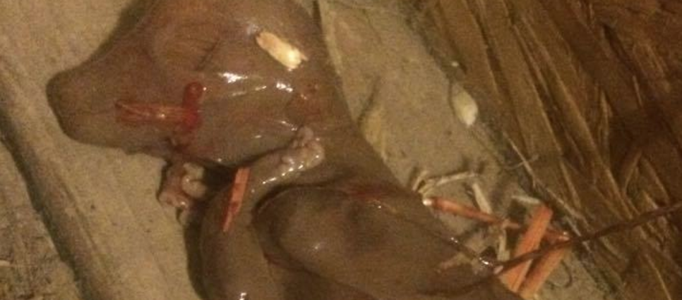
Mummified piglet
Mummified fetus depicting the various stages of the fetal development (photo extracted from maphavet)
Red blood cells in pigs contain nucleus (photo extracted from maphavet)