Not-In-Pig (NIP)

We all get frustrated when our sow/gilt return and we find that they are not in-pig (pregnant). The cause of this is not being vigilant in watching if they return (come back on heat, hogging) or it could be a case of an infection.

Causes

The main causes are poor management and infertility. With regards to management, experience shows us that empty sows and gilts are as of a result of being misidentified poorly, housed in groups running with the boar and services are not supervised and observation of post service for returns to oestrus are not detected. I know it is difficult to keep an eye on our pigs all the time, but if you know when they are due to start hogging then introducing the boar and observing can save you time and worry. Knowing if and when the sow/gilt is due to return by counting 21 days from her last hogging will help you to know if the service has been successful.

If pregnancy testing is not carried out (hire our pregnancy scanner from our website oxfordsandyblackpiggroup.org), then sows may reach term without being pregnant. Infertility may be solely responsible when observation has been a little lax. When embryos are produced and die, sows return to oestrus at uneven intervals. Where disease or cold have reduced the ability of the animal to return or where housing is such that hogging cannot be observed, then the pigs will remain undetected until a routine pregnancy check is followed or the expected farrowing date arrives with no farrowing. Sometimes, in these cases, there may be an undetected abortion, cystic ovaries or pyometra which there will be an infection of the uterine horn.

Clinical signs

‘Not-in-pig’ sows or gilts return to heat after service and also fail to develop the abdominal swelling and underline development typical of pregnancy. The sows are found to be non-pregnant when examined by. A history of vulva discharge may pinpoint the stage at which pregnancy ended.

It will come to no surprise that failure to farrow leads to a diagnosis of not-in-pig. The reasons for the failure of pregnancy can be determined by examining the records. If the recording is not carried out, services are not supervised, pregnancy checks not carried out and returns to oestrus not checked visually, then poor management is the cause.

Pigs that are in poor condition and exposed to too hot, cold, damp and drafty housing then this will result in the sow/gilt not carrying to term and possibly reabsorbing or aborting. Where records confirm that service was carried out and pregnancy has been confirmed reliably, then again, abortion or reabsorption has occurred.

Treatment and prevention

Before animals are treated as not-in-pig, a pregnancy test should be carried out, as the commonest cause of failure to return to service is pregnancy. Cystic ovaries and subdued oestrus can be treated using chorionic gonadotrophin. Pyometra may respond to antimicrobial treatment by injection and allow a return to oestrus.

Prevention is largely a matter of management, although vaccination against conditions such as parvovirus, erysipelas, leptospirosis, PRRS, and influenza can also be a reason for a non-productive sow.

Sows must be identified individually, services must be attended with good recording keeping of the boar or semen used together with results of any pregnancy tests. Ensure that mating is successful and occurs at the correct time if AI-ing that semen quality is adequate. Be vigilant to observe that there are no vulva discharges after service.

To hire our pregnancy scanner, please click here

Mastitis

Most of us have experienced this in our sows and it also affects the gilts. There are a few causes to mastitis such as teat injuries from floors or piglets, bacterial infection, poor hygiene and the symptoms is that the udder is hot, hard and swollen, lumps and sometimes the sow/gilt may become unwell and the piglets may also suffer due to lack of feeding.

Causes

Mastitis in sows is caused by ascending infection of the teats by bacteria. The organism most commonly involved is Escherichia coli or related organisms (coliforms). Bacteria such as Arcanobacterium pyogenes, streptococci and staphylococci may cause infections of single glands. Acute and severe mastitis caused by Klebsiella spp. may occur in outbreaks of fatal disease following trauma to the teats caused by rough sawdust bedding. Traumatic injury resulting from piglet teeth, sawdust bedding, or poor quality flooring add to infection. One or both glands supplying a single teat may be infected. Infection enters via the teat canal following teat contamination and bacteria multiply in the gland. Introduction of endotoxin into the mammary gland will cause mastitis and agalactia and endotoxin can be detected in the blood in mastitis.

Clinical signs

Acutely affected sows are usually depressed, loss or lack of appetite and fevered (temperatures of 40.5-42°C, 105-107°F are not uncommon). The udder is usually swollen and oedematous (abnormal swelling of fluid), often with massive congestion. Any secretion that may be obtained after oxytocin injection contains pus. Pain in the udder may lead to restlessness in the sow when piglets attempt to suck. The litter rapidly loses condition. Acute mastitis usually occurs within 1-3 days of parturition. The body temperature falls below normal, the animal can no longer rise and respiratory distress develops frequently leading to death. Coliform (a bacteria calledaerobic bacillus that lives in the colon) mastitis appears to regress within 3-4 days although in severe cases lactation may cease entirely. Sub-acute infection or infection in one or more glands occurs much more commonly and may be recognised by the increased hardness of the gland and, in its early stages, by a square area of reddening or the skin over the affected gland. The litter loses condition. Mastitis in a single gland is often noted only when an affected gland fails to return to normal after weaning. There is often teat injury, especially in the pairs of teats.

Acute mastitis is easily recognisable as affected sows are off their feed and have obvious swelling of the udder, sometimes with reddening and oedema. The litter is usually in poor condition. Animals are often lying down alot, but must be made to rise in order to confirm that mastitis is present. The udder should be felt on both sides by running the hand under both lines of glands.

Individual affected glands feel firm and hot. Laboratory examination of expressed secretion confirms that presence of mastitis by means of cell counts and the organism(s) responsible can be identified. Action requires oxytocin injection as milk let down is under voluntary control. Sub-clinical mastitis may only be detected after cell counts on expressed milk, showing 75% white blood cells. Chronic mastitis is easily felt upon inspection during lactation and easily seen in dry sows.

Treatment and prevention

Acutely ill sows may be saved by injection with neomycin, tetracyclines, ampicillin, amoxicillin, streptomycin, fluoroquinolone, ceftiofur or trimethoprim: sulphonamide for 2-4 days. Oxytocin should also be given. The litter should be fostered or reared artificially. After recovery it may be necessary to cull the sow as she will be prone to this ailment on all farrowings.

Other supportive treatment may include anti-inflammatory medication. In sub-acute cases, rehydration is not necessary. Antimicrobial injection may improve the condition where single glands are affected, but results are poor where abscess are present and has occurred or teat ends are damaged.

Control depends upon hygiene, use of soft bedding other than sawdust, early treatment and, possibly, the use of commercial E. coli vaccine if the condition is due to E. coli. Where the condition is recurring and pre-farrowing infection is suspected, treatment with trimethoprim sulphonamide at 15 mg/kg given in the feed from day 112 of gestation to day 1 post-partum may eliminate early lesions and prevent the development of clinical disease. Sows which have had severe or repeated bouts of mastitis should, unfortunately, be culled and affected sows should only be retained if they have sufficient functioning teats.

Meningitis

The brain and spinal cord are protected within bony cavities (the skull and the spinal column) but separating the nervous tissue from the bone is a series of membranes called the meninges. When these become inflamed in the condition known as meningitis (usually due to bacterial infection), pressure builds up on the nervous tissue and nervous signs ensue.

In the early stages of meningitis, pigs will be dull and depressed, reluctant to stand and have a raised rectal temperature. Occasionally, they may be seen pressing their head against a wall and they will be unsteady on their legs. As the disease progresses and in response to stimulation (handling, noise), affected pigs will subside into paddling convulsions while lying on their side. Careful observation will reveal that the eyes, when open, will flick from side to side (this is called nystagmus). Death can ensue within a few hours and in some cases of meningitis may simply be found dead.

Infection in the brain results from the bloodborne spread of bacteria, which can gain entry through any break in the skin or mucosa. Sporadic meningitis is particularly seen in the young piglet, where infection gains entry through the navel, tail-dock wound, clipped teeth or fight wounds, and is very much a feature of colostral insufficiency. Bacterial spread in the bloodstream (bacteriaemia) can lead to infection in other areas of the body – particularly the joints, causing joint ill, or arthritis. The bacteria involved are usually environmental contaminants such as staphylococci, E. coli and streptococci. However, epizootic forms of meningitis can occur, particularly in weaner pigs four to ten weeks old and rarely in older growing pigs. The most common cause in these cases is Streptococcus suis type II and Haemophilus parasuis.

Streptococcus suis Meningitis

This is typically a post weaning disease that is triggered by the stress of weaning and by mixing pigs of different ages. Overcrowding, poor ventilation and in particular, high humidity all seem to exacerbate the disease. In an infected herd, the organism is picked up at or soon after birth, the reservoir being the nasal chambers and tonsils as well as the vagina of the sow. The organism colonises the tonsil of the young pig and from there will spread via the bloodstream to the brain.

Onset of the disease can be extremely rapid and sudden death may be seen. Streptococcus suis can readily be cultured from the meninges of an affected pig that has not been treated. Paddling convulsions are a classic feature, along with temperatures of 41degrees C (106f) and above. The disease usually behaves in an all or nothing way – in contrast with most gut or respiratory tract diseases, there is no effect on the growth of unaffected meningitic pigs.

Control can be difficult, vaccines have not proved effective and strategic use of antibiotics such as amoxycillin by injection at weaning may be needed to prevent clinical disease. Medication of weaned pigs via water or feed with penicillin-based antibiotic will often suffice in milder outbreaks. Recovered pigs may drop dead suddenly two or three weeks later, the result of seeding of infection on the heart valves and production of endocarditis.

Worth noting that S. suis type II (along with other strains of S. suis) are zoonotic, so particular care should be taken when handling affected pigs and when attending farrowing in herds known to be infected. Infection for humans is normally by skin penetration through cuts and grazes – thorough hand washing with soap is essential following contact with potentially infect material.

Haemophilus parasuis Meningitis

There are many strains of H. parasuis present within pig populations and, while the most common manifestations of disease are either respiratory or septicaemic, cases do occur where the infection targets the brain, producing meningitis (arthritis is also occasionally seen). The clinical presentation is identical to that described above, but diagnosis can be difficult as the organism is very fragile post mortem – euthanasia of an affected untreated pig and immediate sapling of the meninges or cerebral spinal fluid is essential.

Treatment

Treatment of any form of meningitis is based upon killing the causative organism and providing support therapy. Streptococcus suis is generally very sensitive to antimicrobials treatment with penicillin-based medicines such as amoxycillin, but treatment must be rapid and involve a formulation that achieves high levels of antibiotic in the body immediately. While Haemophilus parasuis is also very sensitive to antibiotics, response to treatment of affected pigs is frequently disappointing, probably due to the fact that clinical signs are the result of toxin release rather than the effect of the bacterium itself; by the time signs are seen, the damage is done and killing off the organism will have no effect.

A common complication of meningitis in pigs is dehydration. Their inability to feed and drink soon leads to fluid shortage, especially in the young pigs. This is often manifests in the form of “salt poisoning” (water-deprivation neuropathy), which can easily be confused with meningitis clinically even though it is quite distinct from it. So many pigs that die through meningitis infection actually subside into salt poisoning, which is the true cause of death.

The pig should be removed from the rest of the litter – where it can be bullied – and placed in a bedded area to prevent injury. Often, response to treatment will be very rapid (within two to four hours). It should be given fluids (water containing electrolytes), preferably by mouth on a little and often basis. Care must be taken to ensure that the pig is swallowing fluids and not inhaling them.

Support treatment with corticosteroids or non-steroidal anti-inflammatory agents can also improve recovery rates. As a general rule, however, a failure to respond to treatment within forty-eight hours is likely to leave the pig permanently brain damaged and so euthanasia is appropriate.

IN ALL CASES PLEASE DISCUSS YOUR SITUATION WITH YOUR VETERINARY PRACTICE.

Photos obtained from Google

Porcine Parvovirus (PPV)

PLEASE REMEMBER TO SIGN UP TO THE DISEASE CHARTER WITH AHDB PORK.

When sourcing stock it is up to you to ask the breeder if they have suffered any diseases within their herd if you don’t ask you don’t know and the breeder will and should not be offended by your question as this shows due diligence on your behalf.

It it my intention to discuss diseases specific to the pregnant sow and the effects her infection will have on the unborn litter and then move on to respiratory disease, nervous system disease etc over the weeks. So hope you find it all helpful.

Reproduction Disease – Porcine Parvovirus (PPV)

Apart from a single very unusual report of skin disease in weaners, PPV is solely associated with the reproductive failure or with its effect on an unborn litter. Infection of the non-pregnant animal has no clinical effect and immunity is acquired that will be life-long and will protect all future litters. The effects that PPV infection will have on a pregnant sow depend on the stage of pregnancy, which I have shown below.

Stage of reproductive cycle Effect of PPV infection Result

Not pregnant No effect Immunity

At service and within Death of fertilised eggs/ Return to service 10 days of service differentiating embryos at 3 weeks 10 – 25 days post service Embryonic death Delayed return to service or small litter 25 – 75 days post-service Foetal death, often Variable-sized mummified progressive through the litter pigs affecting whole or part of the litter, and/or stillbornpigs

75 days plus Minimum effect on foetuses Possibly small pigs born as the immune response can be that have been checked during growth, and stillborn pigs

From this it can be seen that the effects of PPV infection can be stillbirth, mummification, embryonic death and infertility, giving the old acronym SMEDI. It should be noted that abortion is a very rare manifestation of PPV infection.

On a herd basis, in a naïve herd, an outbreak of PPV disease will last two to three months and will manifest by varying signs over that time in the following sequence:

  1. Increased regular returns to oestrus lasting two to three weeks
  2. Increased irregular returns to oestrus lasting two to three weeks simultaneously with above
  3. Stillborn pigs starting simultaneously with above for one to two months.
  4. Increase in mummified pigs from six to twelve weeks after (1) above, and failures to farrow.
  5. Drop in total litter size for two to three weeks from eighty days after the start of the outbreak.

It is a sequential disease, the classic sign of which is large numbers of mummified pigs within a litter, of variable size, starting around one month after an increase in returns to service. The diagnosis of PPV disease is based on clinical pictures supported by blood tests and by virus detection in the livers of mummified or stillborn pigs.

Prevention and Control

Highly effective vaccines against PPV, which we have discussed in the previous post on the group, are available and are given to gilts prior to breeding. The actual programme (number of doses and timing) varies between products, but it should be noted that maternally derived antibodies (those passed to a piglet in colostrums) can survive for up to six months and that these can block vaccine efficacy. Vaccination should, therefore, not be given too early in life. It is also worth noting that the disease can be transmitted from boars to sows or vice versa by direct mating with infected pigs or through artificial insemination from infected semen of boars.

Erysipelas

(When sourcing stock IT IS UP TO YOU to ask the breeder if they have suffered any diseases within their herd if you don’t ask you don’t know and the breeder will and should not be offended by your question as this shows due diligence on your behalf.)

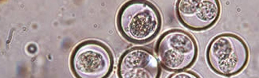
Erysipelas is caused by a bacterium called Erysipelothrix rhusiopathiae in pigs and is one of the oldest recognised diseases that affect growing and adult pigs. The organism commonly resides in the tonsillar tissue. These typical healthy carriers can shed the organism in their faeces or oronasal secretions and are an important source of infection for other pigs.

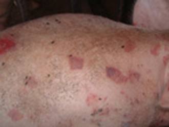
Disease outbreaks may be acute or chronic, and clinically inapparent infections also occur. Acute outbreaks are characterised by sudden and unexpected deaths, febrile episodes, painful joints, and skin lesions that vary from generalized cyanosis to (a bluish discoloration of the skin due to poor circulation or inadequate oxygenation of the blood) the often-described diamond skin (rhomboid urticaria) lesions. Chronic erysipelas tends to follow acute outbreaks and is characterized by enlarged joints and lameness. A second form of chronic erysipelas is vegetative valvular endocarditis. Pigs with valvular lesions may exhibit few clinical signs; however, when exerted physically they may show signs of respiratory distress, lethargy, and cyanosis, and possibly suddenly succumb to the infection.

The genus of Erysipelothrix is subdivided into two major species: E rhusiopathiae and E tonsillarum. In addition, there are other strains that constitute one or more additional species known as E species 1, E species 2, E species 3, and E inopinata. At least 28 different serotypes of Erysipelothrix spp are recognized, and pigs are considered to be susceptible to at least 15. Field cases of swine erysipelas are predominately caused by E rhusiopathiae serotypes 1a, 1b, or 2.

On farms where the organism is endemic, pigs are exposed naturally to E rhusiopathiae when they are young. Maternal-derived antibodies provide passive immunity and suppress clinical disease. Older pigs tend to develop protective active immunity as a result of exposure to the organism, which does not necessarily lead to clinical disease. E rhusiopathiae is excreted by infected pigs in faeces and oronasal secretions, effectively contaminating the environment. When ingested, the organism can survive passage through the hostile environment of the stomach and intestines and may remain viable in the faeces for several months. Recovered pigs and chronically infected pigs may become carriers of E rhusiopathiae. Healthy pigs also may be asymptomatic (showing no symptoms) carriers. Infection is by ingestion of contaminated feed, water, or faeces and through skin abrasions.P

The acute and chronic forms of erysipelas may occur in sequence or separately. Pigs that succumb to the acute septicemic form may die suddenly without previous clinical signs. This form occurs most frequently in growing and finishing pigs. Acutely infected pigs are depressed and reluctant to stand and move. Affected pigs squeal excessively when handled, require assistance to stand, and prefer to lie down soon after being forced to stand. Affected pigs may also walk stiffly on their toes and shift weight from limb to limb when standing. Anorexia and thirst are common, and febrile pigs will often seek wet, cool areas to lie down. Skin discoloration may vary from widespread erythema (reddening of the skin) and purplish discoloration of the ears, snout, and abdomen, to diamond-shaped skin lesions almost anywhere on the body, but particularly on the lateral and dorsal regions. The lesions may occur as discrete, pink or purple areas of varying size that become raised and firm to the touch within 2–3 days of illness. They may disappear over the course of a week or progress to a more chronic type of lesion, commonly referred to as diamond skin disease. If untreated, necrosis and separation of large areas of skin can occur, and the tips of the ears and tail may become necrotic.

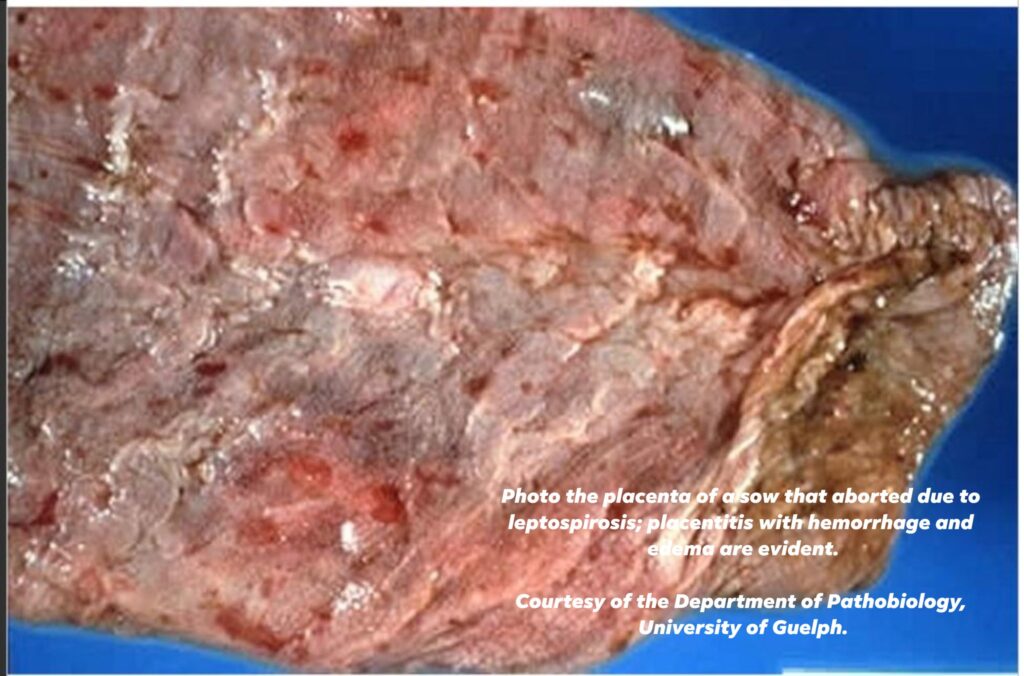
Clinical disease is usually sporadic and affects individuals or small groups, but sometimes larger outbreaks occur. Mortality is variable (0–100%), and death may occur up to 6 days after the first signs of illness. Acutely affected pregnant sows may abort, probably due to the fever, and lactating sows may stop producing milk.

Untreated pigs may develop the chronic form of the disease, usually characterized by chronic arthritis, vegetative valvular endocarditis, or both. Such lesions may also be seen in pigs with no previous signs of septicemia. Valvular endocarditis is most common in mature or young adult pigs and is frequently followed by death, usually from embolism or cardiac insufficiency. Chronic arthritis, the most common form of chronic infection, produces mild to severe lameness. Affected joints may be difficult to detect initially but eventually become hot and painful to the touch and later visibly enlarged. Dark purple, necrotic skin lesions that commonly separates itself from the dead tissue may be seen. Mortality in chronic cases is low, but growth rate is retarded.

Diagnosis of erysipelas is based on clinical signs, gross lesions, response to antimicrobial therapy, and demonstration of the bacterium or DNA in tissues from affected animals. Acute erysipelas can be difficult to diagnose in individual pigs showing only fever, poor appetite, and listlessness. However, in outbreaks involving several animals, the presence of skin lesions and lameness is likely to be seen in at least some cases and would support a clinical diagnosis. Rhomboid urticaria or diamond skin lesions are almost diagnostic when present; however, similar lesions can also be seen with classical swine fever virus.

A rapid, positive response to penicillin therapy in affected pigs supports a diagnosis of acute erysipelas because of the sensitivity of the organism to penicillin.

Chronic erysipelas can be difficult to definitively diagnose. Arthritis and lameness, coupled with the presence of vegetative valvular endocarditis postmortem, may support a presumptive diagnosis of chronic erysipelas.

Serologic tests cannot reliably diagnose erysipelas but can be useful to determine previous exposure or success of vaccination protocols, because antibodies should increase after vaccination.

Treatment:

As we have discussed in previous posts in the group under “Back to Basics Part IV – Handling Medications – What’s in your Cupboard ?” E rhusiopathiae is sensitive to penicillin. Ideally, affected pigs should be treated at 12-hr intervals for a minimum of 3 days, although longer durations of therapy may be necessary to resolve severe infections. On an economic basis, penicillin is the best choice for antibiotic therapy, but ampicillin and ceftiofur also yield satisfactory results in acute cases. When injecting large numbers of affected pigs is impractical, tetracyclines delivered in the feed or water may be useful. Fever associated with acute infections can be managed by administration of NSAIDs such as flunixin meglumine or by delivery of aspirin in the water. Erysipelas antiserum is described as an effective adjunct to antibiotic therapy in treating acute outbreaks but is not commonly available. Treatment of chronic infections is usually ineffective and not cost effective.

Vaccination against E rhusiopathiae is very effective in controlling disease outbreaks.

Optimal timing of vaccination may vary from farm to farm. When E rhusiopathiae is endemic in the production environment, vaccination should precede anticipated outbreaks. Susceptible pigs may be vaccinated before weaning, at weaning, or several weeks after weaning. Male and female pigs selected for addition to the breeding herd should be vaccinated with a booster 3–5 wk later. Thereafter, breeding stock should be vaccinated twice yearly. Vaccines should not be administered to animals undergoing antibiotic therapy, because antibiotics can interfere with the subsequent immune response to the vaccine.

In addition to vaccination, attention to sanitation and hygiene and elimination of pigs with clinical signs suggestive of erysipelas infection represent other viable methods that may help control the disease. But in all cases you must discuss your situation with your vets to discuss your vaccination programme.

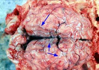
Photos courtesy of Pig333, Dr D Risco Unidad de Patologı´a Infecciosa Photo above showing findings in a lung showing good cells being suffocated by the erysipelas bacterium.

Coccidiosis

Coccidiosis affects all ages of pigs and is a tacky or watery diarrhoea, piglets do not seem to grow very well and can cause death in piglets. It stems from poor hygiene.

Causes

Coccidia are single cell protozoan parasites which multiply in the cells lining the intestine. Cystoisospora suis is the main pathogen of piglets and is responsible for the most coccidial diarrhoea. Cryptosporidium parvum, C. suis and other genotypes may also affect piglets but occurs commonly in weaned pigs and Eimeria species occur in weaned pigs, finishers and gilts but not in piglets. The resistant oocysts of Cystoisospora. suis are eaten and reach the upper small intestine where small sporozoites hatch and invade cells of the intestinal lining within 24 hours. They multiply asexually, damaging their host cell, to give rise to first stage merozoites and then to a further two stages within cells of the small intestinal lining. Each fourth stage cell reaches the lower part of the small intestine at about 4-5 days after infection and invades an epithelial (lining) cell to produce the sexual stage. Some become groups of microgametes and some remain single to become macrogametes. Mating in the gut produces oocysts which appear in the faeces as oocysts within 5 days of infection. Eimeria species multiply in the same way as C. suis, but Cryptosporidium remains in the absorptive, apical part of the cell and appears to prevent absorption of nutrients and causes cell death.

Transmission

Coccidia are passed out in the faeces and Cystoisospora suis oocysts develop within 48 hours at 24-27ºC into resistant hard walled sporulated oocysts which are infective. They are resistant to drying and can persist in piggeries and soil for up to 10 months. The major source of infective oocysts is the faeces of piglets aged between 2 and 4 weeks, when from 4,000 to 400,000 oocysts per gram may be present. Sows may pass a few oocysts and infect their litters, but the major source of both direct and indirect infection is the piglet. Transmission of Eimeria species resembles that of Cystoisospora, although the age range affected differ, but some Cryptosporidium species have more than one host species. The route of infection is oral in all cases.

Signs

Coccidiosis causes mal-absorption, diarrhoea, loss of condition and some deaths. The faeces of affected piglets may be whitish and firm, there may be transient pasty diarrhoea or profuse yellowish, watery diarrhoea between 5-15 days of age and most commonly from 7-10 days of age. Affected piglets may be thinner than unaffected litter mates and remain gaunt and hairy for some weeks thereafter. Severely affected piglets may die. Mortality rates may reach 20%. The diarrhoea responds poorly to antimicrobial therapy and slow growth occurs in recovered animals and diarrhoea rarely continues beyond weaning. Wasting and diarrhoea have been recorded in gilts, finishing pigs and wild boar infected with Eimeria spp. In experimental E. neodebliecki infection, frothy, mucoid diarrhoea occurred 9-12 days post infection. Most infections with C. parvum are asymptomatic (90% in one study). Clinical signs rarely appear before 3 days of age and are most commonly reported in piglets at 10-21 days of age. Affected piglets are depressed and pass a watery, sometimes brownish, diarrhoea, which may persist for 3-5 days. The organism can be recognised in the faeces of diarrhoeic weaners. Affected weaned pigs are often depressed and anorexic, in poor condition with a rough dirty coat and pasty faeces.

Treatment and prevention

Coccidiosis in piglets can be treated with trimethoprim sulphonamide and they should be supported with electrolyte. Toltrazuril given as an oral suspension by stomach tube on day 3 prevents diarrhoea, prevents oocyst shedding and can maintain piglet growth. Scrupulous attention should be given to hygiene in control, as the high levels of oocysts passed by piglets and their ability to survive for months make environmental contamination important. Sows should be cleaned on entry to the farrowing houses which should be disinfected or fumigated with methyl bromide or ammonia (final concentration 2%). Steam cleaning at 65˚C for 15 minutes and proprietary disinfectants can also be used to destroy oocysts. Less disease and oocyst shedding has been demonstrated on fully slatted cleaned floors. Trimethoprim sulphonamide may be given by injection or orally to older pigs with Eimeria species and the inclusion of an anticoccidial such as amprolium at 1 kg permix per tonne in sow feed for 7-10 days prior to farrowing and for 2 weeks afterwards will reduce the shedding of oocysts by sows and other pigs. No treatment has yet been shown to affect Cryptosporidia. The oocysts can be destroyed by the same disinfectants as those of the other coccidia.

Be aware

Cystoisospora and Eimeria infections are of no public health importance, but the pig may be a source of infection with Cryptosporidium for humans and vice versa.

Biotin Deficiency – Nail Growth

This affects all age groups from growers, gilts, sows and boars. It is caused by diet and poor floors and effects Lameness, claw defects, poor growth, diarrhoea, hair loss, scaly skin, small litters.

Causes

Dietary biotin (a water-soluble B vitamin) is normally present in adequate amounts in pig rations, although in feeds containing wheat or barley, the vitamin may be less readily available to pigs than in maize-based diets. Biotin is a cofactor in a number of body enzymes especially in the carboxylation (a chemical reaction in which a carboxylic acid group is produced by treating a substrate with carbon dioxide) or transcarboxylation reactions required for energy metabolism. In growing pigs lipogenesis (fat formation) is affected when the important enzyme, Acetyl CoA carboxylase cannot be formed. Deficiency in biotin on wheat and barley-based rations occurs quite commonly and at least 180 mg/tonne should be present for health.

Mode of transmission

Biotin deficiency is not infectious and occurs where rations are deficient in the vitamin.

Clinical signs

Early changes in biotin deficiency are slight but include progressive hair loss, dry and scaly skin and a white film and transverse grooves on the tongue. After 5-7 weeks on a deficient diet, claw defects occur. Erosion of the heel occurs first and is followed by cracking of the sole. Cracks appear in the now rubbery horn of the sole and the claw wall and result in lameness. Secondary infection may occur. In most cases, foot lesions in sows appear on the underside of the hoof and are only seen when the claw is cleaned. Others appear as longitudinal cracks on the wall originating in the coronary band. Heel and sole erosions and cracks also occur. Inspection of the sows’ feet after thorough cleaning will allow the claw defects to be seen clearly. Cracks arising from the coronary band and erosions of the sole and heel and the presence of alopecia and scaly skin suggest biotin deficiency. As the foot lesions could be mistaken for those of a vesicular disease, such disease should be ruled out. There may be alopecia and a dry scaly skin which may progress to give dermatitis with brownish crusts and pinpoint haemorrhages. The reproductive effects include low numbers born, especially born alive and reared and long weaning to service intervals. Reproductive effects measured over 4 parities indicate at 1-1.4 pigs per sow per year are lost, that weaning to service intervals may be increased by up to 4 days and that conception to the first service may be reduced by 9%. Good recording is essential for this effect to be detected.

Treatment and prevention

A diagnosis of biotin deficiency can be confirmed by analysing the biotin content of the ration (100-220 µg/kg is the normal range) and noting the response to biotin supplementation of the diet. The reproductive effects can be confirmed only by supplementation of the diet in controlled studies. Populations with plasma levels of 60 ng/100 ml benefit from supplementation.

When deficiencies occur it is common practice to supplement the diet with 400 mg-1,250 mg/tonne of D-biotin; it is best to seek the advice of an experienced pig nutritionist. Biotin is available as a 1% d-biotin premix and 40g premix/tonne is necessary for most purposes, for growing gilts, and pregnant and lactating sows to prevent foot lesions and improve litter size. Levels up to 3,000 mg/tonne may be necessary to reverse hoof lesions and time should be allowed for the damaged horn to grow out.

Abortion

This affects both gilts and sows and the causes range from mismanagement, fever, infections of the uterus and poisoning.

Effects: The sow/gilt will abort recognisable foetuses before day 110 of gestation, after which production of dead foetuses is classified as a stillbirth. Sows may be visibly ill before abortion occurs or remain normal.

Causes

Death of the conceptuses may occur early from hormonal causes as four live foetuses are required to maintain an early pregnancy. After implantation, infection is the main cause of abortion, indirectly through fever or directly as a result of the infection of the foetus. Fever is the cause of abortion in erysipelas and other feverish diseases. Invasion of the foetus can occur in many viral and bacterial diseases. In some, foetal invasion results in death and dead foetuses reabsorbed, and in older pregnancies, the foetus may become mummified and later stillbirth may occur. All combinations of normal, resorbing, and mummified foetuses may be aborted. Major infections capable of causing abortion are Aujeszky’s Disease, brucellosis, enterovirus infections, leptospirosis, parvovirus, PRRS, swine fever and toxoplasmosis. Ingestion of the mould toxin zearalenone is also capable of causing abortion, as are management factors, such as high carbon monoxide concentrations.

Mode of transmission

Infectious causes of abortion are transmitted by the routes associated with the causal infection. Non-transmissible abortions occur.

Clinical signs

Sows may be seen to abort and the aborted material may be found on the floor of their accommodation. The sow may not appear ill in some cases (enterovirus, parvovirus, some leptospirosis), but in other diseases such as erysipelas, there is clear evidence that the sow is ill, off feed, and has evidence of the disease. In the case of erysipelas, high fever and the characteristic skin lesions are often found. The rectal temperature should be taken to establish the presence of fever. During or after abortion, the vulva may be bloodstained and the tail and hindquarters may be smeared with sticky discharge. As sows frequently eat aborted material, abortion is hardest to detect in loose-housed animals. Where abortion is missed, the termination of pregnancy may be detected only when an animal, previously known to be pregnant, fails to farrow, returns to oestrus or is identified following a pregnancy check. Abortion is recorded in pig herds as a routine. Abortions should be uncommon in a herd (fewer than 1%), but if they reach 2.5%, the cause should be determined. Management factors may be obvious and in zearalenone poisoning, enlargement of the vulvas of piglets and enlargement of the mammary glands in non-pregnant gilts. The presence of specific diseases may only be identified by laboratory examination of the aborted foetuses, vaginal discharges or blood from the affected sow.

Postmortem lesions

The products of abortion may be visible as embryos (large sacs 10-20 cm in length, with a central dark area, the embryo itself, from which blood vessels radiate) or as perfectly formed pig foetuses. These have their eyes closed and are hairless. They may be partially decayed, mummified or perfectly fresh with haemorrhages on their skins. The age of the foetus when death occurred may be determined by measuring foetal crown-rump length. Placentas may be absent, although they are sometimes passed surrounding aborted piglets. Fluid from the chests of aborted foetuses is used to detect infectious agents by culture or PCR and antibody to them may be present after 70 days gestation. Sows are rarely examined postmortem, but ageing corpora lutea are visible in the ovaries and the uterus may be enlarged, although it rapidly returns to normal. Lesions of a causal infectious disease may be visible but laboratory examination of vulva discharge or uterine contents may be required.

Treatment and prevention

The ability to prevent further abortions varies, depending on the cause. For example, zearalenone toxicity can be eliminated rapidly by feeding clean rations. Carbon monoxide poisoning can be eliminated by adjusting gas heaters. Toxoplasmosis may be prevented by stopping contamination of pig feed by cat faeces. Erysipelas can be treated easily with penicillin and leptospirosis with tetracyclines. Solid immunity follows the elimination of these infections. Less specific infections of the uterus can be eliminated, but ideally, the animals should be culled as persisting infection may reduce fertility.

To control abortion, sows can be vaccinated against erysipelas and parvovirus. In some countries, vaccination against leptospirosis, Aujeszky’s Disease, swine fever and PRRS is also possible. Vaccination requires at least 14 days to protect against an agent and may not protect against venereal infections. There are no vaccines for enteroviruses. Brucellosis is notifiable and controlled by other means. The aborted material should be treated as infectious and correctly discarded, with disinfection as appropriate.

Animals which have aborted should come back into oestrus within 10 days and may then be served. The exception is if body condition has been lost when consideration should be given to increasing feed and delaying service. Those that do not return to oestrus or do not hold to the next service, should, unfortunately, be culled.

Special note

Some infectious causes of abortion (Swine Fever, African Swine Fever, Brucellosis) are notifiable and some, such as Brucellosis and Leptospirosis, can infect humans handling the products of abortion. Please ensure that strict hygiene is practised.